. 2019 Feb;69(2):845-859.
doi: 10.1002/hep.30252. Epub 2019 Jan 3.

NLR Family Pyrin Domain-Containing 3 Inflammasome Activation in Hepatic Stellate Cells Induces Liver Fibrosis in Mice

Affiliations

NLR Family Pyrin Domain-Containing 3 Inflammasome Activation in Hepatic Stellate Cells Induces Liver Fibrosis in Mice

Maria Eugenia Inzaugarat et al. Hepatology. 2019 Feb.

Abstract

The NLR family pyrin domain-containing 3 (NLRP3) inflammasome plays an important role in liver fibrosis (LF) development. However, the mechanisms involved in NLRP3-induced fibrosis are unclear. Our aim was to test the hypothesis that the NLRP3 inflammasome in hepatic stellate cells (HSCs) can directly regulate their activation and contribute to LF. Primary HSCs isolated from wild-type (WT), Nlrp3-/- , or Nlrp3L351PneoR knock-in crossed to inducible (estrogen receptor Cre-CreT) mice were incubated with lipopolysaccharide (LPS) and adenosine triphosphate (ATP), or 4OH-tamoxifen, respectively. HSC-specific Nlrp3L351P knock-in mice were generated by crossing transgenic mice expressing lecithin retinol acyltransferase (Lrat)-driven Cre and maintained on standard rodent chow for 6 months. Mice were then sacrificed; liver tissue and serum were harvested. Nlrp3 inflammasome activation along with HSC phenotype and fibrosis were assessed by RT-PCR, western blotting, fluorescence-activated cell sorting (FACS), enzyme-linked immunosorbent assay, immunofluorescence (IF), and immunohistochemistry (IHC). Stimulated WT HSCs displayed increased levels of NLRP3 inflammasome-induced reactive oxygen species (ROS) production and cathepsin B activity, accompanied by an up-regulation of mRNA and protein levels of fibrotic makers, an effect abrogated in Nlrp3-/- HSCs. Nlrp3L351P CreT HSCs also showed elevated mRNA and protein expression of fibrotic markers 24 hours after inflammasome activation induced with 4-hydroxytamoxifen (4OHT). Protein and mRNA expression levels of fibrotic markers were also found to be increased in isolated HSCs and whole liver tissue from Nlrp3L351P Lrat Cre mice compared to WT. Liver sections from 24-week-old NlrpL351P Lrat Cre mice showed fibrotic changes with increased alpha smooth muscle actin (αSMA) and desmin-positive cells and collagen deposition, independent of inflammatory infiltrates; these changes were also observed after LPS challenge in 8-week-old NlrpL351P Lrat Cre mice. Conclusion: Our results highlight a direct role for the NLRP3 inflammasome in the activation of HSCs directly triggering LF.


Figures

Figure 1:
Figure 1:. Induction of canonical NLRP3 inflammasome activation accompanied by ROS production and cathepsin B in hepatic stellate cells by LPS/ATP.
Cultured HSC from WT mice, but not from Nlrp3−/−, display augmented protein and mRNA expression levels for Nlrp3 and pro-Il1b as well as mature IL-1β (supernatant) after LPS and/or LPS/ATP stimulation (A-B). Flow cytometry analysis showed that WT HSC increase caspase1 activity after LPS/ATP stimulation, which was not seen in Nlrp3−/− HSC (C). mRNA levels of Casp11, Il1a and Hmgb1, effectors of non-canonical activation of NLRP3, are not affected by LPS or LPS/ATP stimulation (D). LPS triggers ROS production in WT HSC, evidenced by increased DCF fluorescence, which was further enhanced after subsequent ATP stimulation, an effect not present in Nlrp3−/− HSC (E). Increased cathepsin B was detected in WT HSC after LPS/ATP stimulation. Interestingly, Nlrp3−/− HSC showed reduced cathepsin B levels compared to WT HSC. (6 replicates were included for all measured values, * = p<0.05, ** = p<0.01)
Figure 2:
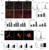
Figure 2:. Effect of NLRP3 inflammasome activation in hepatic stellate cells fibrotic phenotype
After LPS/ATP stimulation, HSC from WT but not Nlrp3−/− mice showed an increase in collagen 1 and αSMA expression along with a change in the shape (A). Flow cytometry analysis showed that WT HSC displayed increased vimentin levels after LPS/ATP stimulation, an effect not present in Nlrp3−/− HSC (B). mRNA levels of Ctgf, aSma, Tgfb, Vim, and Coll1a1 were found to be increased after LPS, and further enhanced after subsequent ATP stimulation (C). HSC isolated from Nlrp3L351P/+ CreT showed a pronounced change in shape along with increased collagen 1 and αSMA expression (D), augmented vimentin expression measured by FACS (E) and elevated mRNA levels of aSma, Coll1a1, Vim, Ctgf and Tgfb (F) after NLRP3 inflammasome overactivation was induced by 4-OH-tamoxifen, effects that were intensified after the addition of LPS. (6 replicates were included for all measured values, * = p<0.05, ** = p<0.01)
Figure 3:
Figure 3:. Result of Lrat Cre-driven expression of mutant gain-of-function NLRP3 on hepatic stellate cell phenotype
Western blot of whole liver lysates showed augmented Nlrp3 inflammasome-related proteins in Nlrp3L351P/+ Lrat Cre mice (A), supported by quantification of protein levels of NLRP3, Caspase1 and IL-1β (B) as well as elevated mRNA levels of pro-Il1b and pro-Casp1 (C). Immunofluorescence showed increased protein expression of αSMA and desmin in isolated HSC from Nlrp3L351P/+ Lrat Cre mice (D). Further analysis in whole liver lysates confirmed increased protein expression (E, F) as well as increased mRNA levels (F) of aSma in Nlrp3L351P/+ Lrat Cre mice compared to control littermates. (n= 8 mice per group for all measured values, * = p<0.05, ** = p<0.01)
Figure 4:
Figure 4:. Effect of persistent NLRP3 activation in hepatic stellate cells on liver fibrosis
Representative pictures of αSMA, desmin and Sirius Red stained liver sections from Nlrp3L351P Lrat Cre and control animal littermates (A). Quantification of stained sections showed an increased number of αSMA (B) and desmin (C) positive cells in Nlrp3L351P Lrat compared to littermates. Staging of fibrosis showed the presence of mild predominantly perisinusoidal fibrosis (D) in Nlrp3L351P/+ Lrat Cre mice accompanied by an upregulation of mRNA levels of Col1a1, Col3a1 and Timp1 (E) compared to littermate control animals. Increased hydroxyproline concentration in whole liver lysates of Nlrp3L351P/+ Lrat Cre mice compared to littermates (F). (n=3–5 mice per group for all measured values, * = p<0.05, ** = p<0.01)
Figure 5:
Figure 5:. Inflammation and liver injury after persistent NLRP3 activation in hepatic stellate cells
Representative pictures of H&E, TUNEL staining (A), F4/80 and MPO (C) stained liver sections from Nlrp3L351P Lrat Cre and control littermates. Serum ALT levels were unchanged in Nlrp3L351P/+ Lrat Cre compared to littermate mice (B). Quantification of immunohistochemistry showed no detectable changes in F4/80 (D) or MPO (E) positive cells between Nlrp3L351P/+ Lrat Cre and the littermate control mice. (n=5 mice per group for all measured values, * = p<0.05, ** = p<0.01)
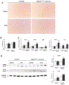
Figure 6.
Figure 6.
Representative pictures of Sirius Red stained liver sections from LPS or Saline injected Nlrp3L351P/+ Lrat Cre and littermates (A). Sirius red staining quantification showed no changes in collagen deposition (B). Nevertheless, LPS-injected Nlrp3L351P/+ Lrat Cre displayed an upregulation of mRNA levels of Ctgf, Col1a1, Tgfb, aSma and Vim (E) compared to saline and LPS injected littermates. Increased αSMA and CTGF protein concentration in whole liver lysates of Nlrp3L351P/+ Lrat Cre mice compared to littermates (D). (n=4–6 mice per group for all measured values, * = p<0.05 vs. WT saline, # = p<0.01 vs. WT LPS)
Figure 7:
Figure 7:
Summary of the role of Nlrp3 inflammasome in HSC activation and liver fibrosis development

References

    1. Strowig T, Henao-Mejia J, Elinav E, Flavell R. Inflammasomes in health and disease. Nature 2012;481:278–286. - PubMed
    1. Wen H, Ting JP, O’Neill LA. A role for the NLRP3 inflammasome in metabolic diseases--did Warburg miss inflammation? Nat Immunol 2012;13:352–357. - PMC - PubMed
    1. Gombault A, Baron L, Couillin I. ATP release and purinergic signaling in NLRP3 inflammasome activation. Front Immunol 2012;3:414. - PMC - PubMed
    1. Gross CJ, Mishra R, Schneider KS, Medard G, Wettmarshausen J, Dittlein DC, Shi H, et al. K(+) Efflux-Independent NLRP3 Inflammasome Activation by Small Molecules Targeting Mitochondria. Immunity 2016;45:761–773. - PubMed
    1. He Y, Hara H, Nunez G. Mechanism and Regulation of NLRP3 Inflammasome Activation. Trends Biochem Sci 2016;41:1012–1021. - PMC - PubMed

Publication types