Image quality and dose characteristics for an O-arm intraoperative imaging system with model-based image reconstruction
- PMID: 30180274
- PMCID: PMC6711149
- DOI: 10.1002/mp.13167
Image quality and dose characteristics for an O-arm intraoperative imaging system with model-based image reconstruction
Abstract
Purpose: To assess the imaging performance and radiation dose characteristics of the O-arm CBCT imaging system (Medtronic Inc., Littleton MA) and demonstrate the potential for improved image quality and reduced dose via model-based image reconstruction (MBIR).
Methods: Two main studies were performed to investigate previously unreported characteristics of the O-arm system. First is an investigation of dose and 3D image quality achieved with filtered back-projection (FBP) - including enhancements in geometric calibration, handling of lateral truncation and detector saturation, and incorporation of an isotropic apodization filter. Second is implementation of an MBIR algorithm based on Huber-penalized likelihood estimation (PLH) and investigation of image quality improvement at reduced dose. Each study involved measurements in quantitative phantoms as a basis for analysis of contrast-to-noise ratio and spatial resolution as well as imaging of a human cadaver to test the findings under realistic imaging conditions.
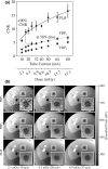
Results: View-dependent calibration of system geometry improved the accuracy of reconstruction as quantified by the full-width at half maximum of the point-spread function - from 0.80 to 0.65 mm - and yielded subtle but perceptible improvement in high-contrast detail of bone (e.g., temporal bone). Standard technique protocols for the head and body imparted absorbed dose of 16 and 18 mGy, respectively. For low-to-medium contrast (<100 HU) imaging at fixed spatial resolution (1.3 mm edge-spread function) and fixed dose (6.7 mGy), PLH improved CNR over FBP by +48% in the head and +35% in the body. Evaluation at different dose levels demonstrated 30% increase in CNR at 62% of the dose in the head and 90% increase in CNR at 50% dose in the body.
Conclusions: A variety of improvements in FBP implementation (geometric calibration, truncation and saturation effects, and isotropic apodization) offer the potential for improved image quality and reduced radiation dose on the O-arm system. Further gains are possible with MBIR, including improved soft-tissue visualization, low-dose imaging protocols, and extension to methods that naturally incorporate prior information of patient anatomy and/or surgical instrumentation.
Keywords: cone-beam CT; image-guided surgery; model-based image reconstruction; radiation dose; surgical navigation.
© 2018 American Association of Physicists in Medicine.
Conflict of interest statement
Author PH is an employee of Medtronic.
Figures


Similar articles
-
A mobile isocentric C-arm for intraoperative cone-beam CT: Technical assessment of dose and 3D imaging performance.Med Phys. 2020 Mar;47(3):958-974. doi: 10.1002/mp.13983. Epub 2020 Jan 6. Med Phys. 2020. PMID: 31863480 Free PMC article.
-
Mobile C-arm cone-beam CT for guidance of spine surgery: image quality, radiation dose, and integration with interventional guidance.Med Phys. 2011 Aug;38(8):4563-74. doi: 10.1118/1.3597566. Med Phys. 2011. PMID: 21928628 Free PMC article.
-
Intraoperative cone-beam and slot-beam CT: 3D image quality and dose with a slot collimator on the O-arm imaging system.Med Phys. 2021 Nov;48(11):6800-6809. doi: 10.1002/mp.15221. Epub 2021 Sep 23. Med Phys. 2021. PMID: 34519364 Free PMC article.
-
Known-component 3D image reconstruction for improved intraoperative imaging in spine surgery: A clinical pilot study.Med Phys. 2019 Aug;46(8):3483-3495. doi: 10.1002/mp.13652. Epub 2019 Jun 30. Med Phys. 2019. PMID: 31180586 Free PMC article.
-
AAPM Task Group Report 238: 3D C-arms with volumetric imaging capability.Med Phys. 2023 Aug;50(8):e904-e945. doi: 10.1002/mp.16245. Epub 2023 Feb 20. Med Phys. 2023. PMID: 36710257 Free PMC article. Review.
Cited by
-
Cone-Beam CT image contrast and attenuation-map linearity improvement (CALI) for brain stereotactic radiosurgery procedures.J Appl Clin Med Phys. 2018 Nov;19(6):200-208. doi: 10.1002/acm2.12477. Epub 2018 Oct 19. J Appl Clin Med Phys. 2018. PMID: 30338919 Free PMC article.
-
Known-component metal artifact reduction (KC-MAR) for cone-beam CT.Phys Med Biol. 2019 Aug 21;64(16):165021. doi: 10.1088/1361-6560/ab3036. Phys Med Biol. 2019. PMID: 31287092 Free PMC article.
-
Feasibility of a method for low contrast CT image quality assessment using difference detail curves for abdominal scans.Z Med Phys. 2022 May;32(2):209-217. doi: 10.1016/j.zemedi.2022.01.001. Epub 2022 Feb 17. Z Med Phys. 2022. PMID: 35184974 Free PMC article. German.
-
Three-dimensional intraoperative computed tomography imaging for zygomatic fracture repair.J Korean Assoc Oral Maxillofac Surg. 2021 Oct 31;47(5):382-387. doi: 10.5125/jkaoms.2021.47.5.382. J Korean Assoc Oral Maxillofac Surg. 2021. PMID: 34713813 Free PMC article.
-
Radiation dose and image quality comparison during spine surgery with two different, intraoperative 3D imaging navigation systems.J Appl Clin Med Phys. 2019 Feb;20(2):136-145. doi: 10.1002/acm2.12534. Epub 2019 Jan 24. J Appl Clin Med Phys. 2019. PMID: 30677233 Free PMC article.
References
-
- Siewerdsen JH, Moseley DJ, Burch S, et al. Volume CT with a flat‐panel detector on a mobile, isocentric C‐arm: pre‐clinical investigation in guidance of minimally invasive surgery. Med Phys. 2005;32:241–254. - PubMed
-
- Guerrero ME, Jacobs R, Loubele M, Schutyser F, Suetens P, van Steenberghe D. State‐of‐the‐art on cone beam CT imaging for preoperative planning of implant placement. Clin Oral Investig. 2006;10:1–7. - PubMed
-
- Gupta R, Cheung AC, Bartling SH, et al. Flat‐panel volume CT: fundamental principles, technology, and applications. Radiographics. 2008;28:2009–2022. - PubMed
-
- Houten JK, Nasser R, Baxi N. Clinical assessment of percutaneous lumbar pedicle screw placement using theO‐arm multidimensional surgical imaging system. Neurosurgery. 2012;70:990–995. - PubMed
-
- Helm PA, Teichman R, Hartmann SL, Simon D. Spinal navigation and imaging: history, trends, and future. IEEE Trans Med Imaging. 2015;34:1738–1746. - PubMed
MeSH terms
Grants and funding
LinkOut - more resources
Full Text Sources
Other Literature Sources
Miscellaneous

