

Different sulfonylureas induce the apoptosis of proximal tubular epithelial cell differently via closing KATP channel
- PMID: 30180807
- PMCID: PMC6122448
- DOI: 10.1186/s10020-018-0042-5
Different sulfonylureas induce the apoptosis of proximal tubular epithelial cell differently via closing KATP channel
Abstract
Background: Sulfonylureas (SUs) are widely prescribed for the treatment of type 2 diabetes (T2DM). Sulfonylurea receptors (SURs) are their main functional receptors. These receptors are also found in kidney, especially the tubular cells. However, the effects of SUs on renal proximal tubular epithelial cells (PTECs) were unclear.
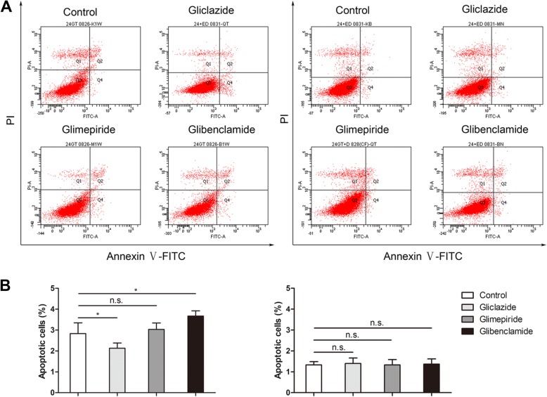
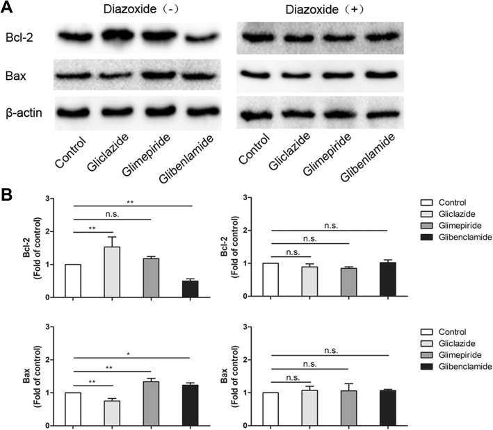
Methods: Three commonly used SUs were included in this study to investigate if different SUs have different effects on the apoptosis of PTECs. HK-2 cells were exposed to SUs for 24 h prior to exposure to 30 mM glucose, the apoptosis rate was evaluated by Annexin/PI flow cytometry. Bcl-2, Bax and the ratio of LC3II to LC3I were also studied by western blot in vitro. Diazoxide was used to evaluate the role of KATP channel in SUs-induced apoptosis of PTECs. A Student's t-test was used to assess significance for data within two groups.
Results: Treatment with glibenclamide aggravated the apoptosis of HK-2 cells in high-glucose, as indicated by a significant decrease in the expression of Bcl-2 and increase in Bax. Additionally, the decreased LC3II/LC3I reflects that the autophagy was inhibited by glibenclamide. Similar but less pronounced change was found in glimepiride group, however, nearly opposite effects were found in gliclazide group. Further, the effects of glibenclamide on apoptosis promotion and the decreased LC3II/LC3I were ameliorated obviously by treatment with 100uM diazoxide. The potential protection effect of gliclazide was also inhibited after opening the KATP channel.
Conclusion: Our results suggest that, the effects of glibenclamide and glimepiride on PTECs apoptosis, especially the former, were achieved in part by closing the KATP channel. In contrast to glibenclamide and glimepiride, therapeutic concentrations of gliclazide showed an inhibitory effect on apoptosis of PTECs, which may have a benefit in the preservation of functional PTECs mass.
Keywords: ATP-dependent potassium channel; Diabetes kidney disease; Glibenclamide; Gliclazide; Glimepiride; Proximal tubular epithelial cells.
Conflict of interest statement
Ethics approval and consent to participate
Not applicable
Consent for publication
Not applicable
Competing interests
The authors declare that they have no competing interests.
Publisher’s Note
Springer Nature remains neutral with regard to jurisdictional claims in published maps and institutional affiliations.
Figures

Similar articles
-
Comparative Effects of Three Sulfonylureas (Glibenclamide, Glimepiride, and Gliclazide) on Proliferation and Migration of Vascular Smooth Muscle Cells.Cell Physiol Biochem. 2019;52(1):16-26. doi: 10.33594/000000002. Epub 2019 Feb 18. Cell Physiol Biochem. 2019. PMID: 30790502
-
Effects of sulfonylureas on K(ATP) channel-dependent vasodilation.J Diabetes Complications. 2003 Mar-Apr;17(2 Suppl):6-10. doi: 10.1016/s1056-8727(02)00273-8. J Diabetes Complications. 2003. PMID: 12623162
-
Differential effects of sulphonylureas on the vasodilatory response evoked by K(ATP) channel openers.Fundam Clin Pharmacol. 2003 Feb;17(1):61-9. doi: 10.1046/j.1472-8206.2003.00144.x. Fundam Clin Pharmacol. 2003. PMID: 12588631
-
Characterization of the molecular mode of action of the sulfonylurea, glimepiride, at beta-cells.Horm Metab Res. 1996 Sep;28(9):464-8. doi: 10.1055/s-2007-979838. Horm Metab Res. 1996. PMID: 8911984 Review.
-
The molecular interaction of sulfonylureas with beta-cell ATP-sensitive K(+)-channels.Diabetes Res Clin Pract. 1995 Aug;28 Suppl:S67-80. doi: 10.1016/0168-8227(95)01076-p. Diabetes Res Clin Pract. 1995. PMID: 8529521 Review.
Cited by
-
A High-Fat Diet Increases Activation of the Glucagon-Like Peptide-1-Producing Neurons in the Nucleus Tractus Solitarii: an Effect that is Partially Reversed by Drugs Normalizing Glycemia.Cell Mol Neurobiol. 2022 Aug;42(6):1995-2002. doi: 10.1007/s10571-021-01079-2. Epub 2021 Apr 3. Cell Mol Neurobiol. 2022. PMID: 33811589 Free PMC article.
-
Manoalide Induces Intrinsic Apoptosis by Oxidative Stress and Mitochondrial Dysfunction in Human Osteosarcoma Cells.Antioxidants (Basel). 2023 Jul 14;12(7):1422. doi: 10.3390/antiox12071422. Antioxidants (Basel). 2023. PMID: 37507960 Free PMC article.
-
The Functional Interaction of KATP and BK Channels with Aquaporin-4 in the U87 Glioblastoma Cell.Biomedicines. 2024 Aug 19;12(8):1891. doi: 10.3390/biomedicines12081891. Biomedicines. 2024. PMID: 39200356 Free PMC article.
-
A High-Content Screen for the Identification of Plant Extracts with Insulin Secretion-Modulating Activity.Pharmaceuticals (Basel). 2021 Aug 17;14(8):809. doi: 10.3390/ph14080809. Pharmaceuticals (Basel). 2021. PMID: 34451906 Free PMC article.
-
Role of renal tubular programed cell death in diabetic kidney disease.Diabetes Metab Res Rev. 2023 Feb;39(2):e3596. doi: 10.1002/dmrr.3596. Epub 2022 Nov 30. Diabetes Metab Res Rev. 2023. PMID: 36401596 Free PMC article. Review.
References
Publication types
MeSH terms
Substances
LinkOut - more resources
Full Text Sources
Other Literature Sources
Research Materials
Miscellaneous
