. 2018 Sep 4;14(1):14.
doi: 10.1186/s13005-018-0171-7.

Bilateral Central Giant Cell Granuloma of the mandibular angle in three females from the same family

Affiliations

Bilateral Central Giant Cell Granuloma of the mandibular angle in three females from the same family

Simona Tecco et al. Head Face Med. .

Abstract

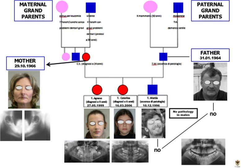
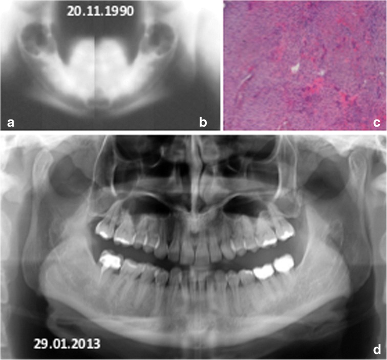
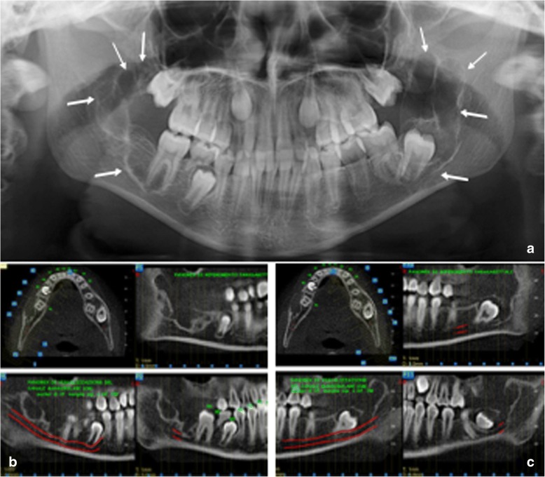
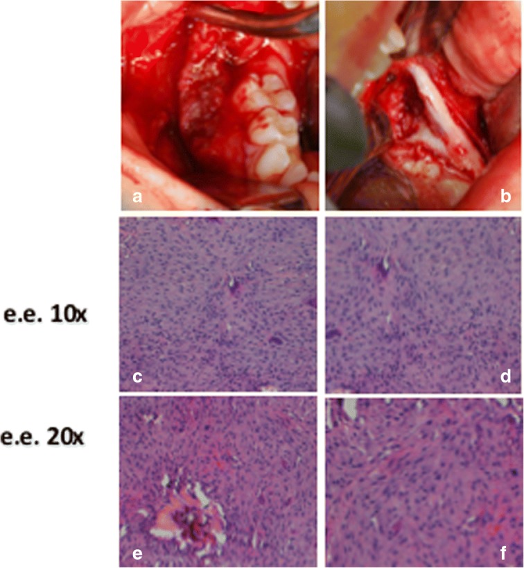
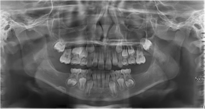
In literature there are few reports about multiple CGCG. But this is the first report of bilateral CGCG of the mandibular angles in three females from the same family.This report describes three cases of females from the same family - a mother and two young daughters - with bilateral CGCG in their jaw angles. All the lesions were surgically removed and the histopathologic diagnosis was always identical: giant cell central granulomas, with patterns that were absolutely superimposable between them and with that of the mother.The hypothesis is that this presentation of CGCG may be defined as hereditary bilateral CGCG of the mandibular angles (or also, cherubism-like lesions).

Keywords: Case series; Central Giant cells granuloma; Cherubism-like lesions.


Conflict of interest statement

Ethics approval and consent to participate

Ethics approval was obtained by the Ethic Committee of the University of L’Aquila, Italy. The consent to the treatment was obtained by the patients before the beginning of the therapy.

The partecipants have signed consent to the surgical intervention, the processing of personal data and the use of clinical material for scientific purposes (C.F.Uni.L’AquilaHosp.S.S.1995 and C.F.Uni.L’AquilaHosp.S.S.2012).

Consent for publication

The consent to publish the present data was obtained from the subjects, also for the children. The partecipants have signed consent to the surgical intervention, the processing of personal data and the use of clinical material for scientific purposes (C.F.Uni.L’AquilaHosp.S.S.1995 and C.F.Uni.L’AquilaHosp.S.S.2012).

Competing interests

The authors declare that they have no competing interests.

Publisher’s Note

Springer Nature remains neutral with regard to jurisdictional claims in published maps and institutional affiliations.

Figures

Fig. 1
Fig. 1
The family
Fig. 2
Fig. 2
Mother: diagnosis at age 23. Mandibular x-Ray tomography: the right ramus (a) and the left ramus (b) show bilateral and symmetric radiolucenct areas. c Histopathological pattern suggests central giant cell granuloma. d Panoramic radiography 23 years after surgery; note the complete restoring of the mandibular bone structure
Fig. 3
Fig. 3
TA, female, diagnosis at age 9 (4-gen-2008) - a Panoramic radiography shows on both side of the mandible two symmetric large multilocular radiolucent lesions involving the angle and the ramus regions (white arrows). In the lower dental arch, there are only the first molar at right side (b) and the first and second molars at left side (c) CTCB study of the mandible, respectively, of the right and the left site, shows the extension of the lesions. Note their critical relationship with the mandibular canal and its neurovascular structures, in particular the inferior alveolar nerves
Fig. 4
Fig. 4
TA, female, diagnosis at age 9 – a-c and d-f: respectively the lesion of the right mandibular side and the left mandibular side. Note for each one the intraoperative aspect, and e.e. 10× and e.e. 20× histopatological speciments that show a moderately cellular and partially collagenized stroma, characterized by melted cells with dense nuclei and giant cells osteoclast like
Fig. 5
Fig. 5
TA, female, diagnosis at age 9 – Follow-up at 5 years (16-avr-2013). Panoramic radiography shows the good aspect of the bone mandibular structures and the absence of relapse
Fig. 6
Fig. 6
TC, female, diagnosis at age 6 (29-mar-2012) - a Panoramic radiography shows on both side of the mandible two symmetric large multilocular radiolucent lesions involving the angle and the ramus regions and the second molars. (white arrows). In the lower dental arch there are the first and the second molars (b, c) CTCB study of the mandible, respectively, of the right and the left site, shows the extension of the lesions. Note their critical relationship with the mandibular canal and its neurovascular structures, in particular the inferior alveolar nerves
Fig. 7
Fig. 7
TA, female, diagnosis at age 9 – a-d and e-h: respectively the lesion of the right mandibular side and the left mandibular side. Note for each one the intraoperative aspect, the excised tissue, e.e. 10× and e.e. 20× histopatological speciments that show a moderately cellular and partially collagenized stroma, characterized by melted cells with dense nuclei and giant cells osteoclast like
Fig. 8
Fig. 8
Absolute correspondence of the histological aspect of the lesions in the two sisters: the histological framework consisted of a moderately cellular and partially collagenized stroma, characterized by melted cells with dense nuclei and giant cells osteoclast like
Fig. 9
Fig. 9
TA, female, diagnosis at age 9. Follow-up at 8 months years (16-oct-2013). Panoramic radiography shows the good aspect of the bone mandibular structures and the absence of relapse

Similar articles

Cited by

References

    1. Barnes L, Eveson JW, Reichart P. Sidransky D (eds.): World Health Organization classification of tumors. Pathology genetics of head and neck tumors. Lyon: IARC Press; 2005.
    1. Orhan E, Erol S, Deren O, Sevin A, Ekici O, Erdoğan B. Idiopathic bilateral central giant cell reparative granuloma of jaws: a case report and literature review. Int J Pediatr Otorhinolaryngol. 2010;74:547–552. doi: 10.1016/j.ijporl.2010.02.006. - DOI - PubMed
    1. Chrcanovic BR, Gomez RS, Freire-Maia B. Neurofibromatosis type 1 associated with bilateral central giant cell granuloma of the mandible. J Craniomaxillofac Surg. 2011;39:538–543. doi: 10.1016/j.jcms.2010.10.014. - DOI - PubMed
    1. Edwards PC, Fox J, Fantasia JE, Goldberg J, Kelsch RD. Bilateral central giant cell granulomas of the mandible in an 8-year-old girl with Noonan syndrome (Noonan-like/multiple giant cell lesion syndrome) Oral Surg Oral Med Oral Pathol Oral Radiol Endod. 2005;99:334–340. doi: 10.1016/j.tripleo.2004.08.021. - DOI - PubMed
    1. Saccucci M, D'Attilio M, Rodolfino D, Festa F, Polimeni A, Tecco S. Condylar volume and condylar area in class I, class II and class III young adult subjects. Head Face Med. 2012;8:34. doi: 10.1186/1746-160X-8-34. - DOI - PMC - PubMed

MeSH terms

LinkOut - more resources