. 2018 Sep 4;37(1):215.
doi: 10.1186/s13046-018-0890-4.

PHF8 upregulation contributes to autophagic degradation of E-cadherin, epithelial-mesenchymal transition and metastasis in hepatocellular carcinoma

Affiliations

PHF8 upregulation contributes to autophagic degradation of E-cadherin, epithelial-mesenchymal transition and metastasis in hepatocellular carcinoma

Wuhua Zhou et al. J Exp Clin Cancer Res. .

Erratum in

Abstract

Background: Plant homeodomain finger protein 8 (PHF8) serves an activator of epithelial-mesenchymal transition (EMT) and is implicated in various tumors. However, little is known about PHF8 roles in hepatocellular carcinoma (HCC) and regulating E-cadherin expression.

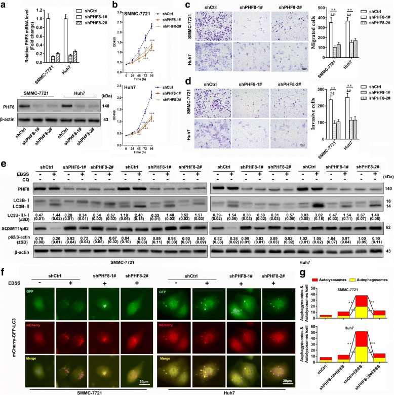
Methods: PHF8 expression pattern was investigated by informatic analysis and verified by RT-qPCR and immunochemistry in HCC tissues and cell lines. CCK8, xenograft tumor model, transwell assay, and tandem mCherry-GFP-LC3 fusion protein assay were utilized to assess the effects of PHF8 on proliferation, metastasis and autophagy of HCC cells in vitro and in vivo. ChIP, immunoblot analysis, rescue experiments and inhibitor treatment were used to clarify the mechanism by which PHF8 facilitated EMT, metastasis and autophagy.

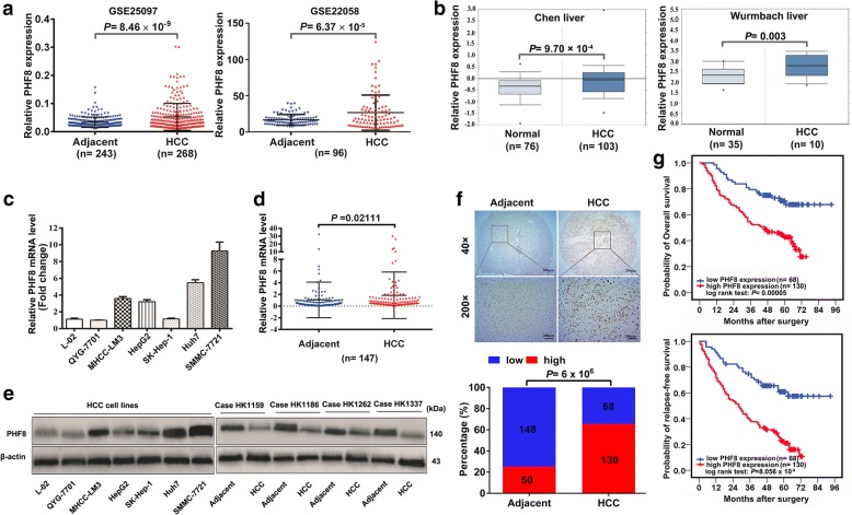
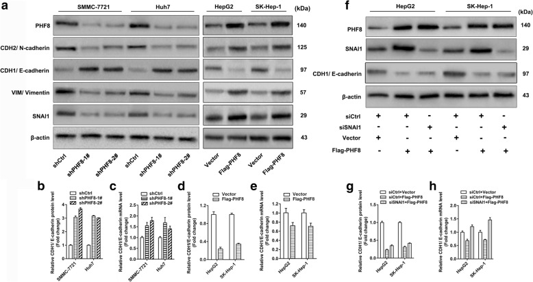
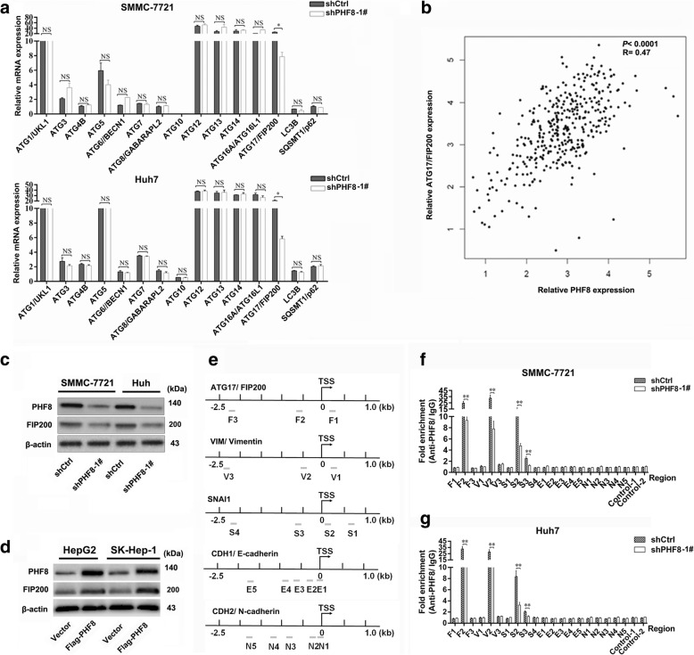
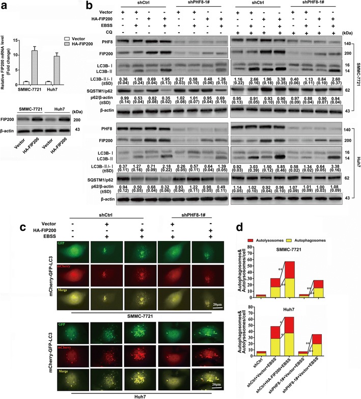
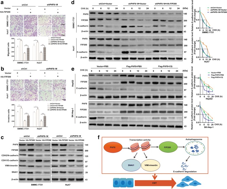
Results: PHF8 upregulation was quite prevalent in HCC tissues and closely correlated with worse overall survival and disease-relapse free survival. Furthermore, PHF8-knockdown dramatically suppressed cell growth, migration, invasion and autophagy, and the expression of SNAI1, VIM, N-cadherin and FIP200, and increased E-cadherin level, while PHF8-overexpression led to the opposite results. Additionally, FIP200 augmentation reversed the inhibited effects of PHF8-siliencing on tumor migration, invasion and autophagy. Mechanistically, PHF8 was involved in transcriptionally regulating the expression of SNAI1, VIM and FIP200, rather than N-cadherin and E-cadherin. Noticeably, E-cadherin degradation could be accelerated by PHF8-mediated FIP200-dependent autophagy, a crucial pathway complementary to transcriptional repression of E-cadherin by SNAI1 activation.

Conclusion: These findings suggested that PHF8 played an oncogenic role in facilitating FIP200-dependent autophagic degradation of E-cadherin, EMT and metastasis in HCC. PHF8 might be a promising target for prevention, treatment and prognostic prediction of HCC.

Keywords: Autophagy; Epithelial-mesenchymal transition, EMT; Hepatocellular carcinoma, HCC; Metastasis; Plant homeodomain finger protein 8, PHF8.


Conflict of interest statement

Ethics approval and consent to participate

The study protocol was approved by the Research Ethics Committee of The First Affiliated Hospital, Zhejiang University, and written informed content was obtained from each enrolled patient. Animal study was initially approved by Animal Care and Use Committee of Zhejiang University, and conducted under the National Institute Guide for the Care and Use of Laboratory Animals.

Consent for publication

Not applicable.

Competing interests

The authors declare that they have no competing interests.

Publisher’s Note

Springer Nature remains neutral with regard to jurisdictional claims in published maps and institutional affiliations.

Figures

Fig. 1
Fig. 1
PHF8 expression is prevalently upregulated and indicated a poor prognosis in HCC. a, b Comparison of PHF8 expression in HCC tissues and normal liver tissues or adjacent normal liver tissues according to the analysis of data from GEO database (GSE25097 and GSE22058), and Oncomine database (Chen liver and Wurmbach liver). c, d Relative PHF8 mRNA level in HCC cell lines and normal human hepatocytes, and in HCC tissues and adjacent normal liver tissues by qRT-PCR analysis. e PHF8 protein expression in HCC cell lines and HCC tissues and adjacent normal tissues by western-blot analysis. β-actin was used as the loading control. f Representative immunohistochemical staining for PHF8 (upper panel, magnification, × 40, × 200) and the percentages of low or high PHF8 expression in paired HCC samples (lower panel). g Kaplan-Meier analysis of overall survival and relapse-free survival of HCC patients with low (n = 68) and high (n = 130) expression of PHF8 based on IHC scoring. Data were presented as mean ± SD
Fig. 2
Fig. 2
PHF8 knockdown significantly suppresses proliferation, migration, invasion and autophagy of HCC cells in vitro. a Determination of transfection efficiency of shRNAs targeting PHF8 in SMMC-7721 and Huh7 cells by qRT-PCR and western-blot assay. Scramble shRNA (shCtrl) was used for negative control. b Inhibited proliferation of SMMC-7721 and Huh7 cells in PHF8 knockdown group by CCK8 assasy (n = 6). c, d Representative images and quantification of migrated and invasive cells by transwell assay in SMMC-7721 and Huh7 cells (n = 3, magnification, × 100). e Representative immunoblot result of autophagy markers, LC3B and p62 in SMMC-7721 and Huh7 cells with PHF8 knockdown. Both cell lines transfected with indicated shRNAs were cultured in complete medium with 10% FBS or EBSS starvation condition with or without CQ (100 μmol) for 8-h. The ratio of LC3-II to LC3-I and p62 to β-actin were shown at the bottom of each band (n = 3). f Representative fluorescence images of autophagosomes and autolysosomes in SMMC-7721 and Huh7 cells with PHF8 knockdown by tandem mCherry-GFP-LC3 fusion protein assay (magnification, × 400). g Quantification of autophagosomes and autolysosomes from random 5 high-power fields of the merged images of each group. * P < 0.05, ** P < 0.01, *** P < 0.001. Data were presented by mean ± SD
Fig. 3
Fig. 3
PHF8 regulates the expression of EMT markers. a Western-blot analysis of expression of SNAI1, VIM/ Vimentin, CDH2/ N-cadherin and E-cadherin. b, d quantification of E-cadherin protein amount. c, e qRT-PCR analysis of E-cadherin mRNA expression in SMMC-7721 and Huh7 cells with PHF8 knockdown and HepG2 and SK-Hep-1 cells with PHF8 exogenous overexpression. f-h Analysis of E-cadherin protein and mRNA expression after SNAI1 knockdown by siRNA in HepG2 and SK-Hep-1 cells with PHF8 overexpression. siCtrl was used for negative control. Data were presented by mean ± SD from three independent experiments
Fig. 4
Fig. 4
PHF8 is involved in transcriptional activation of FIP200, SNAI1 and VIM. a Effect of PHF8-silencing on mRNA expression of key autophagy-related genes. b The relationship between PHF8 expression and FIP200 expression by Gene Expression Profiling Interactive Analysis (GEPIA, http://gepia.cancer-pku.cn/). c, d Western-blot analysis of FIP200 expression in SMMC-7721 and Huh7 cells with PHF8 knockdown and HepG2 and SK-Hep-1 cells with PHF8 exogenous overexpression. e Schematic illustration of the sites of primer amplicons on promoter regions of FIP200, VIM, SNAI1, CDH1 and CDH2 for ChIP analysis. f, g Combining ChIP and qRT-PCR analysis to measure the levels of PHF8 presence at promoter of FIP200, VIM, SNAI1, CDH1 and CDH2 in SMMC-7721 and Huh7 cells with PHF8 knockdown. * P < 0.05, ** P < 0.01, NS, no significance. Data were presented by mean ± SD from three independent experiments
Fig. 5
Fig. 5
FIP200 of exogenous overexpression reverses the effect of PHF8-silencing on autophagy. a qRT-PCR and western-blot analysis of FIP200 expression in SMMC-7721 and Huh7 cells with exogenous overexpression of FIP200. Vector represented empty plasmid for negative control. b Representative immunoblot result of LC3B and p62 in SMMC-7721 and Huh7 cells with co-transfection of indicated shRNAs and plasmids (Vector or HA-FIP200) after cultured in complete medium with 10% FBS or EBSS starvation condition with or without CQ (100 μmol) for 8-h. c Representative fluorescence images of autophagosomes and autolysosomes in SMMC-7721 and Huh7 cells with co-transfection of shRNAs (shCtrl or shPHF8) and plasmids (Vector or HA-FIP200) (magnification, × 400). d Quantification of autophagosomes and autolysosomes from random 5 high-power fields of the merged images of each group. * P < 0.05, ** P < 0.01. Data were presented by mean ± SD from three independent experiments
Fig. 6
Fig. 6
PHF8 promotes EMT and metastasis partly dependent on FIP200-mediated autophagic E-cadherin degradation. a, b Exogenous overexpression of FIP200 reverses effects of PHF8-knockdown on migration and invasion of SMMC-7721 and Huh7 cells (magnification, × 100). c Western-blot analysis of expression of FIP200 and EMT markers in SMMC-7721 and Huh7 cells with co-transfection of shPHF8–1# and HA-FIP200. d, e Western-blot analysis of E-cadherin expression in SMMC-7721 and Huh7 cells with co-transfection of shRNAs (shCtrl or shPHF8–1#) and plasmids (Vector or HA-FIP200), and in HepG2 and SK-Hep-1 cells transfected with PHF8 overexpression and pre-treated by CQ (100 μmol) for 12-h, and subsequently treated by CHX (20 μmol) for indicated time. The protein amount of PHF8 and FIP200 were measured as well. f Schematic representation of the mechanism of PHF8 promotes metastasis by transcriptional regulating expression of SNAI1 and VIM and FIP200-dependent autophagic degradation of E-cadherin. Data were presented as mean ± SD. * P < 0.05, ** P < 0.01

Similar articles

Cited by

References

    1. Siegel RL, Miller KD, Jemal A. Cancer statistics, 2017. Ca A Cancer J Clin. 2017;67:7–30. doi: 10.3322/caac.21387. - DOI - PubMed
    1. Heimbach J, Kulik LM, Finn R, Sirlin CB, Abecassis M, Roberts LR, Zhu A, Murad MH, Marrero J. Aasld guidelines for the treatment of hepatocellular carcinoma. Hepatology. 2017;67:358–380. doi: 10.1002/hep.29086. - DOI - PubMed
    1. Dhir M, Melin AA, Douaiher J, Lin C, Zhen WK, Hussain SM, Geschwind JF, Doyle MB, Abou-Alfa GK, Are C. A review and update of treatment options and controversies in the Management of Hepatocellular Carcinoma. Ann Surg. 2016;263:1112–1125. doi: 10.1097/SLA.0000000000001556. - DOI - PubMed
    1. Serrano-Gomez SJ, Maziveyi M, Alahari SK. Regulation of epithelial-mesenchymal transition through epigenetic and post-translational modifications. Mol Cancer. 2016;15:18. doi: 10.1186/s12943-016-0502-x. - DOI - PMC - PubMed
    1. Smith BN, Bhowmick NA. Role of EMT in metastasis and therapy resistance. J Clin Med. 2016;5:17. doi: 10.3390/jcm5020017. - DOI - PMC - PubMed

MeSH terms