Review
. 2018 Sep 4;10(9):a034801.
doi: 10.1101/cshperspect.a034801.

Protocells and RNA Self-Replication

Affiliations
Review

Protocells and RNA Self-Replication

Gerald F Joyce et al. Cold Spring Harb Perspect Biol. .

Abstract

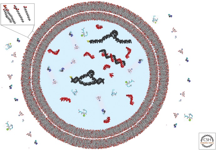
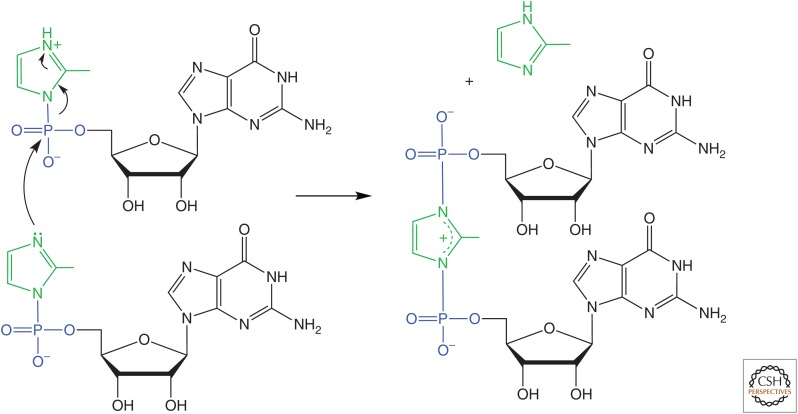
The general notion of an "RNA world" is that, in the early development of life on the Earth, genetic continuity was assured by the replication of RNA, and RNA molecules were the chief agents of catalytic function. Assuming that all of the components of RNA were available in some prebiotic locale, these components could have assembled into activated nucleotides that condensed to form RNA polymers, setting the stage for the chemical replication of polynucleotides through RNA-templated RNA polymerization. If a sufficient diversity of RNAs could be copied with reasonable rate and fidelity, then Darwinian evolution would begin with RNAs that facilitated their own reproduction enjoying a selective advantage. The concept of a "protocell" refers to a compartment where replication of the primitive genetic material took place and where primitive catalysts gave rise to products that accumulated locally for the benefit of the replicating cellular entity. Replication of both the protocell and its encapsulated genetic material would have enabled natural selection to operate based on the differential fitness of competing cellular entities, ultimately giving rise to modern cellular life.


Figures

Figure 1.
Figure 1.
Schematic diagram of a protocell. Protocells bounded by multiple bilayer membranes composed of simple amphiphilic molecules would have been permeable to nucleotides and metal ions complexed with citrate or other ligands. Larger genomic and functional RNA molecules would have been trapped within the protocell interior. Protocell replication would involve growth and division of the membrane boundary as well as replication of the genomic RNA.
Figure 2.
Figure 2.
Potential prebiotic synthesis of pyrimidine nucleosides. (A) Reaction of ribose with cyanamide to form a bicyclic product, with cyanamide joined at both the anomeric carbon and 2-hydroxyl. (B) Reaction of glycolaldehyde with cyanamide in neutral phosphate buffer, followed by addition of glyceraldehyde, to form ribose and arabinose amino-oxazoline (and lesser amounts of the xylose and lyxose compounds). Arabinose amino-oxazoline then reacts with cyanoacetylene to give cytosine 2′,3′-cyclic phosphate as the major product. (C) Analogous reaction of arabinose-3-phosphate to form a bicyclic product, which then reacts with cyanoacetylene to form a tricyclic intermediate that hydrolyzes to give a mixture of cytosine arabinoside-3′-phosphate and cytosine 2′,3′-cyclic phosphate.
Figure 3.
Figure 3.
Potential prebiotic synthesis of 2-thio-C starting from ribose amino-oxazoline (RAO). RAO, which is generated by the reaction of glyceraldehyde with 2-amino-oxazole, crystallizes readily, leading to its accumulation as a reservoir of purified material. Subsequent reaction with cyanoacetylene generates an anhydronucleoside intermediate, which upon attack by hydrosulfide gives the α-anomer of 2-thio-C. Exposure to ultraviolet light results in photoanomerization to give the β-anomer of 2-thio-C, which then desulfurizes to form cytosine.
Figure 4.
Figure 4.
The structures of (A) RNA; (B) p-RNA; (C) TNA; and (D) GNA.
Figure 5.
Figure 5.
Formation of a 5′,5′-linked imidazolium-bridged dinucleotide, which is the reactive intermediate in template-directed polymerization of imidazole-activated nucleotides.
Figure 6.
Figure 6.
X-ray crystal structure of the (A) L1 ligase, and (B) class I ligase ribozymes. Insets show the putative magnesium ion-binding sites at the respective ligation junctions. The structures are rendered in rainbow continuum, with the 5′-triphosphate-bearing end of the ribozyme colored violet and the 3′-hydroxyl-bearing end of the substrate colored red. The phosphate at the ligation junction is shown in white, and the proximate magnesium ion (modeled for the class I ligase) is shown as a yellow sphere, with dashed lines indicating coordination contacts.

References

    1. Adamala K, Szostak JW. 2013a. Nonenzymatic template-directed RNA synthesis inside model protocells. Science 342: 1098–1100. - PMC - PubMed
    1. Adamala K, Szostak JW. 2013b. Competition between model protocells driven by an encapsulated catalyst. Nat Chem 5: 495–501. - PMC - PubMed
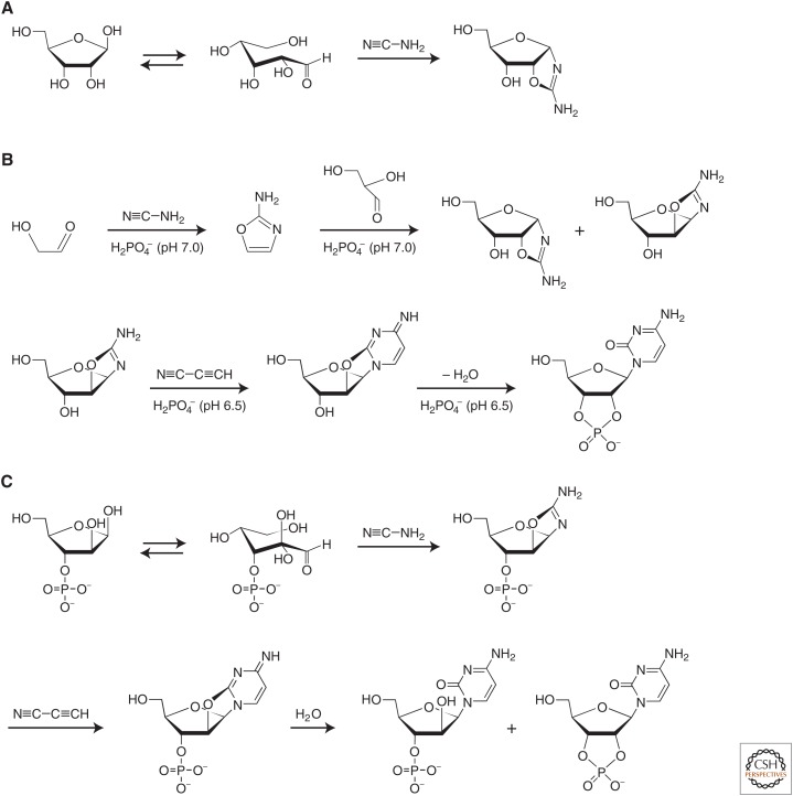
    1. Anastasi C, Crowe MA, Sutherland JD. 2007. Two-step potentially prebiotic synthesis of α-d-cytidine-5′-phosphate from d-glyceraldehyde-3-phosphate. J Am Chem Soc 129: 24–25. - PubMed
    1. Attwater J, Wochner A, Holliger P. 2013. In-ice evolution of RNA polymerase ribozyme activity. Nat Chem 5: 1011–1018. - PMC - PubMed
    1. Aumiller WM Jr, Pir Cakmak F, Davis BW, Keating CD. 2016. RNA-based coacervates as a model for membraneless organelles: Formation, properties, and interfacial liposome assembly. Langmuir 32: 10042–10053. - PubMed

Publication types

LinkOut - more resources