. 2018 Oct 19;293(42):16402-16412.
doi: 10.1074/jbc.RA118.004567. Epub 2018 Sep 4.

Interplay of ancestral non-primate lentiviruses with the virus-restricting SAMHD1 proteins of their hosts

Affiliations

Interplay of ancestral non-primate lentiviruses with the virus-restricting SAMHD1 proteins of their hosts

Sarah A Mereby et al. J Biol Chem. .

Abstract

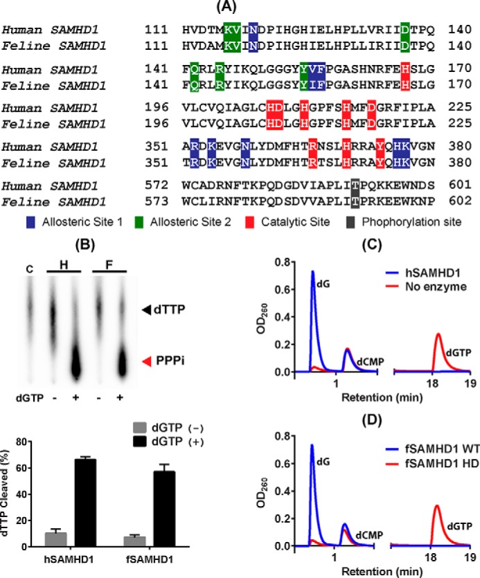
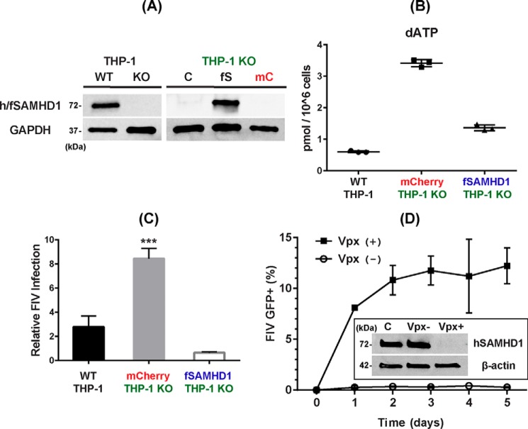
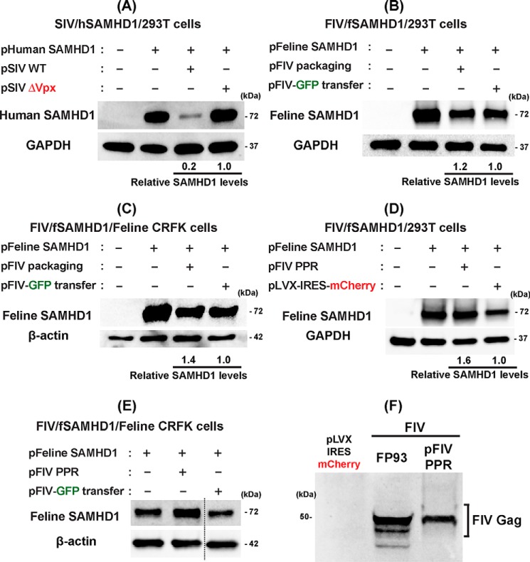
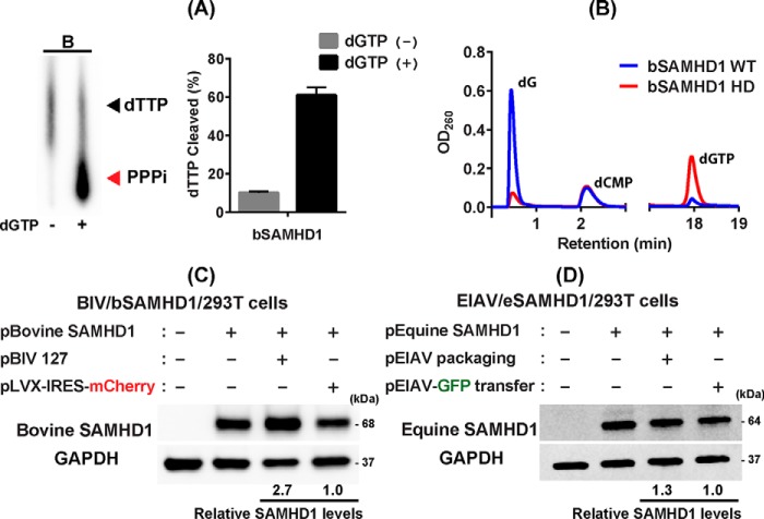
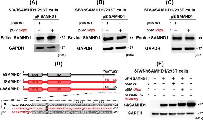
Lentiviruses infect both dividing CD4+ T cells and nondividing myeloid cells, and the infected myeloid cells serve as long-living viral reservoirs. Host sterile alpha motif- and histidine-aspartate domain-containing protein 1 (SAMHD1) kinetically restricts reverse transcription of primate lentiviruses, including human immunodeficiency virus, type 1 (HIV-1) and simian immunodeficiency virus (SIV), in nondividing myeloid cells. SAMHD1 enforces this restriction through its dNTP triphosphohydrolase (dNTPase) activity that depletes cellular dNTPs. Some primate lentiviruses, such as HIV-2, SIVsm, and SIVagm, counteract SAMHD1 restriction by using their viral accessory proteins (Vpx or Vpr) that induce the proteosomal degradation of SAMHD1 and increase dNTP levels. SAMHD1 is conserved among non-primate mammals such as cats, cows, and horses that also carry their own lentiviruses (feline and bovine immunodeficiency viruses and equine infectious anemia viruses, respectively). However, whether these viruses also target SAMHD1 is unknown. Here, we tested whether these ancestral non-primate lentiviruses also can counteract their host SAMHD1 proteins by promoting their proteosomal degradation. Using biochemical and various cell-based assays, we observed that SAMHD1 proteins from the non-primate host species display dGTP-dependent dNTPase activity, but that the non-primate lentiviruses fail to proteosomally degrade the SAMHD1 proteins of their hosts. Our findings suggest that accessory protein-mediated proteosomal degradation of SAMHD1 did not exist among the ancestral non-primate lentiviruses and was uniquely gained by some primate lentiviruses after their transmission to primate species.

Keywords: SAM domain and HD domain-containing protein 1 (SAMHD1); antiviral defense; dNTPase; immunodeficiency virus; lentivirus; macrophage; nucleotide; proteosomal degradation; reverse transcription.


Conflict of interest statement

The authors declare that they have no conflicts of interest with the contents of this article

Figures

Figure 1.
Figure 1.
dNTP hydrolysis activity of feline SAMHD1 protein. A, sequence comparison between human and feline SAMHD1 proteins in HD active site, two allosteric sites (A1 and A2), and C-T phosphorylation site. Red residues, histidine/aspartate residues and residues known to be important for the dNTPase catalytic activity; blue residues, A1 site interacting with dGTP/GTP regulator; green residues, A2 site interacting with dNTPs; and black residue, threonine phosphorylation site (Thr-592 in hSAMHD1). Numbers indicate the first and last amino acid positions of the specified regions for each protein. NCBI reference sequences NM_015474.3 for hSAMHD1 and XM_003983547.2 for fSAMHD1. B, TLC-based assay for dGTP-dependent dTTP hydrolysis. [α-32P]dTTP substrate was incubated with 1 μg purified WT hSAMHD1 (H) and fSAMHD1 (F) in the presence (+) and absence (−) of dGTP for 30 min at 37 °C and heat-killed for 10 min at 70 °C, which was analyzed by TLC (32). PPPi, triphosphate product. Percentages of the cleaved dTTP substrate obtained from triplicated assays were plotted with the error bars representing S.D. C, HPLC-based dNTPase assay for hSAMHD1. dGTP was incubated with (blue line) and without (red line) hSAMHD1 protein for 30 min at 37 °C, and the dG product was detected after separation from dGTP substrate by HPLC. dCMP was added to each reaction as an internal loading control. D, HPLC-based dNTPase assay for fSAMHD1 protein. dGTP was incubated with WT fSAMHD1 (blue line) and fSAMHD1 HD mutant protein (red line). This is a representative data set from triplicates.
Figure 2.
Figure 2.
Cellular dNTP reduction activity and antiviral activity of fSAMHD1 in nondividing THP-1 macrophages. A, expression of fSAMHD1 in hSAMHD1 KO THP-1 cells. Left, human SAMHD1 expression in the PMA-treated parental THP-1 macrophages (WT) was previously abolished by CRISPR/Cas9 (KO) (33). hSAMHD1 expression was detected by Western blotting with anti-SAMHD1 antibody in the differentiated THP-1 cells. Cellular GAPDH protein was used as a loading control. Right, hSAMHD1 KO THP-1 cells (THP-1 KO) were transduced with lentiviral vector co-expressing both HA-tagged fSAMHD1 (fS) and mCherry protein or lentiviral vector expressing only mCherry protein (mC), and the mCherry+ cells were FACS-sorted. The sorted mCherry+ THP-1 were then differentiated to macrophages by PMA for 7 days and analyzed for the fSAMHD1 expression (fS) by Western blotting with HA antibody. GAPDH was used as a loading control. C, untransduced SAMHD1 KO THP-1 control cells. B, cellular dNTP reduction by fSAMHD1 expression in nondividing hSAMHD1 KO THP-1 macrophages. The parental THP-1 cells expressing hSAMHD1 (WT THP-1), hSAMHD1 KO THP-1 cells expressing only mCherry protein (mCherry THP-1 KO), and hSAMHD1 KO THP-1 cells expressing both fSAMHD1 and mCherry protein (fSAMHD1 THP-1 KO) were differentiated by PMA for 7 days, and the dNTP levels in these cells were determined by the RT-based dNTP assay. The total dNTP amounts were normalized by 1 × 10⁶ cells. dATP levels are shown in this figure, and other three dNTP data are in Fig. S2. C, FIV restriction by fSAMHD1 in nondividing hSAMHD1 KO THP-1 macrophages. The parental THP-1 cells expressing hSAMHD1 (WT THP-1), hSAMHD1 KO THP-1 cells expressing only mCherry protein (mCherry THP-1 KO), and hSAMHD1 KO THP-1 cells expressing HA-tagged fSAMHD1 were differentiated by PMA, and transduced with an equal amount of FIV-GFP vector with error bars representing S.D. ***, p ≤ 0.001, using two-tailed Student's t test. The -fold changes of the GFP+ cells compared with the untransduced THP-1 negative background control cells (ratio = 1) in triplicates were plotted. The transduction efficiencies of the vectors used (% GFP ± S.D.) were 1.187 ± 0.384, 3.567 ± 0.361, and 0.813 ± 0.101 for WT THP-1, mCherry THP-1 KO, and fSAMHD1 THP-1 KO, respectively. D, FIV restriction by hSAMHD1 in human primary monocyte-derived macrophages. Human primary monocyte-derived macrophages prepared from four healthy donors were pretreated with VLPs with (Vpx+) or without Vpx (Vpx−) for 12 h, and then transduced with an equal amount of FIV-GFP vector. The transduced cells were collected every day for 5 days, and the percent of the GFP+ cells in triplicates was determined by FACS. Insert shows Western blotting conducted to confirm the degradation of hSAMHD1 by Vpx in human primary macrophages: β-actin was used as a loading control, and antiSAMHD1 antibody was used for detecting hSAMHD1 in the human primary macrophages. C, no VLP pretreatment control macrophages.
Figure 3.
Figure 3.
Test for fSAMHD1 degradation by FIV. A, degradation of hSAMHD1 by SIV. Human 293T cells were co-transfected with pLVX-IRES-mCherry co-expressing HA-tagged hSAMHD1 and mCherry protein (pHuman SAMHD1) (0.1 μg) and a plasmid expressing SIVmac251 proteins either with (pSIV WT) (2 μg) or without Vpx (pSIV ΔVpx) (2 μg), and the hSAMHD1 levels were determined by Western blotting with anti-hSAMHD1 antibody. GAPDH was used as a loading control. Transfection efficiency for the cells expressing hSAMHD1 were monitored by the mCherry protein expression. B, test for the degradation of fSAMHD1 by FIV. 293T cells were co-transfected with a plasmid co-expressing HA-tagged fSAMHD1 and mCherry (pFeline SAMHD1) (0.1 μg) and either FIV packaging plasmid expressing FIV protein except Env protein, FP93 (pFeline packaging) (2 μg) (34) or FIV transfer plasmid expressing only GFP, pGINSIN FIV-GFP (pFIV-GFP transfer) (2 μg). fSAMHD1 levels were visualized by HA antibody. Transfection efficiency was monitored by the mCherry expression. C, test for the degradation of fSAMHD1 by FIV in feline CRFK cells. Feline CRFK cells were co-transfected with the fSAMHD1 expressing plasmid and either FP93 packaging plasmid or pGINSIN FIV-GFP transfer plasmid as described in B. The fSAMHD1 expression was visualized by HA antibody, and cellular β-actin was used as a loading control. D, test for the degradation of fSAMHD1 by infectious molecular clone of pFIV-PPR. The experiment described in (B) was repeated except using full-length FIV-PPR molecular clone. pLVX-IRES-mCherry was used as a negative control. E, the experiment described in (C) was repeated except using full-length FIV-PPR molecular clone. The fSAMHD1 expression was visualized by HA antibody, and cellular β-actin was used as a loading control. The data presented in this figure are representative data from more than three independent transfections, and mean relative SAMHD1 levels shown were calculated by densitometry analysis. The calculated mean ± S.D. values are (A) 0.21 ± 0.05, (B) 1.21 ± 0.49, (C) 1.40 ± 0.57, and (D) 1.60 ± 0.2. F, detection of FIV Gag protein in the 293T cells transfected with FIV plasmids. The same lysates prepared from 293T cells transfected with FP93 and pFIV-PPR used in (B) pFIV packaging and in (D) pFIV PPR were used to detect FIV Gag protein expression with anti-FIV gag antibody. Cells transfected with pLVX-IRES-mCherry expressing only mCherry protein were used as a negative control.
Figure 4.
Figure 4.
Test for bSAMHD1 degradation by BIV and eSAMHD1 degradation by EIAV. A and B, TLC-based (A) and HPLC-based (B) dNTPase activity assays of bSAMHD1 protein. Both TLC- and HPLC-based dNTPase assay were conducted with purified bSAMHD1 protein. These assays were performed in triplicates as described for fSAMHD1 in Fig. 1. [α-32P]dTTP and dGTP were used as SAMHD1 substrates in TLC- and HPLC-based assays, respectively. PPPi, triphosphate product. The HPLC data show the dGTP hydrolysis and dG product by WT bSAMHD1 (blue line) and HD mutant (red line). dCMP is the internal loading control. C, test for degradation of bSAMHD1 by BIV. Human 293T cells were co-transfected with pLVX-IRES-mCherry co-expressing HA-tagged bSAMHD1 and mCherry protein (pBovine SAMHD1) (0.1 μg) and either a full-length molecular clone of BIV (pBIV 127) (2 μg), or pLVX-IRES-mCherry expressing on mCherry protein (pLVX-IRES-mCherry) (2 μg). The bSAMHD1 protein levels were determined in Western blotting with HA antibody. GAPDH was used as a loading control. D, test for degradation of eSAMHD1 by EIAV. Human 293T cells were co-transfected with pLVX-IRES-mCherry co-expressing HA-tagged eSAMHD1 and mCherry protein (pEquine SAMHD1) (0.1 μg) and either EIAV vector packaging plasmid (pEIAV packaging) (2 μg) or GFP-expressing EIAV vector transfer plasmid (pEIAV-GFP transfer) (2 μg). The eSAMHD1 protein levels were determined by Western blotting with HA antibody. GAPDH was used as a loading control. The Western blotting data presented in this figure are representative data from more than three independent transfections, and mean relative SAMHD1 levels shown were calculated by densitometry analysis. The calculated mean ± S.D. values are (C) 2.69 ± 0.40 and (D) 1.29 ± 0.27.
Figure 5.
Figure 5.
Test for non–primate host SAMHD1 protein degradation by Vpx and fSAMHD1 C-T region for Vpx recognition. A–C, tests for Vpx-induced degradation of the non-primate host SAMHD1 proteins. 293T cells were transfected with pLVX-IRES-mCherry co-expressing HA-tagged SAMHD1 proteins from the three non-primate host species (A, fSAMHD1; B, bSAMHD1; and C, eSAMHD1) (0.1 μg) and either pSIV WT (2 μg) or pSIV ΔVpx (2 μg), and the SAMHD1 protein levels were determined by Western blotting with HA antibody. GAPDH was used as a loading control. D, construct of fSAMHD1 chimeric construct (f-hSAMHD1) containing the C-terminal Vpx recognition region of hSAMHD1. The sequence of the hSAMHD1 C-terminal region recognized by Vpx as well as the sequence of fSAMHD1 at the equivalent region is in dotted box. Asterisk sites are the positions of hSAMHD1 depicted for the Vpx recognition (PDB ID: 4CC9). The numbers indicate the amino acid positions of each SAMHD1. The f-hSAMHD1 chimera was constructed by swapping the fSAMHD1 C terminus (residues 586–627) with hSAMHD1 C terminus (residues 585–626) containing Vpx recognition region. The C-term phosphorylation sites (Thr-592 and Thr-593 in human and feline SAMHD1, respectively) are shaded in dark gray for reference. E, test for the degradation of f-hSAMHD1 chimeric construct by SIV. 293T cells were co-transfected with pLVX-IRES-mCherry co-expressing HA-tagged f-hSAMHD1 hybrid construct and mCherry protein (pF-H SAMHD1) (0.1 μg) and either pSIV WT (2 μg) or pSIV ΔVpx (2 μg). pLVX-IRES-mCherry expressing only mCherry protein (2 μg) was used as a negative control. The f-hSAMHD1 chimeric protein was detected by HA antibody. The Western blotting data presented in this figure are representative data from more than three independent transfections.

References

    1. Weissman D., Rabin R. L., Arthos J., Rubbert A., Dybul M., Swofford R., Venkatesan S., Farber J. M., and Fauci A. S. (1997) Macrophage-tropic HIV and SIV envelope proteins induce a signal through the CCR5 chemokine receptor. Nature 389, 981–985 10.1038/40173 - DOI - PubMed
    1. Lewis P. F., and Emerman M. (1994) Passage through mitosis is required for oncoretroviruses but not for the human immunodeficiency virus. J. Virol. 68, 510–516 - PMC - PubMed
    1. Swanstrom A. E., Del Prete G. Q., Deleage C., Elser S. E., Lackner A. A., and Hoxie J. A. (2018) The SIV envelope glycoprotein, viral tropism, and pathogenesis: Novel insights from nonhuman primate models of AIDS. Curr. HIV Res. 16, 29–40 10.2174/1570162X15666171124123116 - DOI - PubMed
    1. Weinberg J. B., Matthews T. J., Cullen B. R., and Malim M. H. (1991) Productive human immunodeficiency virus type 1 (HIV-1) infection of nonproliferating human monocytes. J. Exp. Med. 174, 1477–1482 10.1084/jem.174.6.1477 - DOI - PMC - PubMed
    1. Williams K. C., and Hickey W. F. (2002) Central nervous system damage, monocytes and macrophages, and neurological disorders in AIDS. Annu. Rev. Neurosci. 25, 537–562 10.1146/annurev.neuro.25.112701.142822 - DOI - PubMed

Publication types

Substances

Associated data