

The chaperonin TRiC/CCT is essential for the action of bacterial glycosylating protein toxins like Clostridium difficile toxins A and B
- PMID: 30181275
- PMCID: PMC6156611
- DOI: 10.1073/pnas.1807658115
The chaperonin TRiC/CCT is essential for the action of bacterial glycosylating protein toxins like Clostridium difficile toxins A and B
Abstract
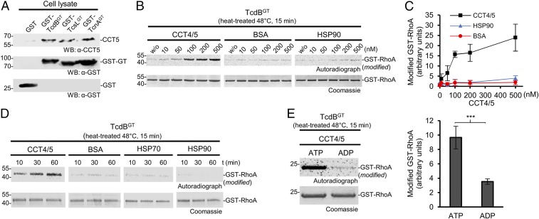
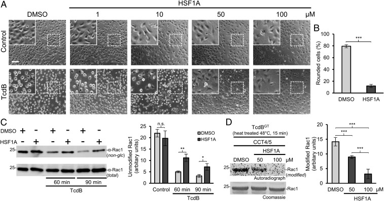
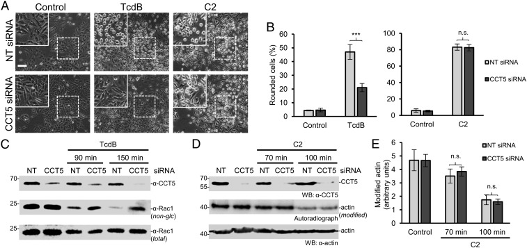
Various bacterial protein toxins, including Clostridium difficile toxins A (TcdA) and B (TcdB), attack intracellular target proteins of host cells by glucosylation. After receptor binding and endocytosis, the toxins are translocated into the cytosol, where they modify target proteins (e.g., Rho proteins). Here we report that the activity of translocated glucosylating toxins depends on the chaperonin TRiC/CCT. The chaperonin subunits CCT4/5 directly interact with the toxins and enhance the refolding and restoration of the glucosyltransferase activities of toxins after heat treatment. Knockdown of CCT5 by siRNA and HSF1A, an inhibitor of TRiC/CCT, blocks the cytotoxic effects of TcdA and TcdB. In contrast, HSP90, which is involved in the translocation and uptake of ADP ribosylating toxins, is not involved in uptake of the glucosylating toxins. We show that the actions of numerous glycosylating toxins from various toxin types and different species depend on TRiC/CCT. Our data indicate that the TRiC/CCT chaperonin system is specifically involved in toxin uptake and essential for the action of various glucosylating protein toxins acting intracellularly on target proteins.
Keywords: Clostridium difficile toxins; TRiC/CCT; chaperonin; glycosyltransferase toxins; toxin uptake.
Conflict of interest statement
The authors declare no conflict of interest.
Figures

Similar articles
-
The Chaperonin TRiC/CCT Inhibitor HSF1A Protects Cells from Intoxication with Pertussis Toxin.Toxins (Basel). 2024 Jan 10;16(1):36. doi: 10.3390/toxins16010036. Toxins (Basel). 2024. PMID: 38251252 Free PMC article.
-
Clostridium difficile Toxin Biology.Annu Rev Microbiol. 2017 Sep 8;71:281-307. doi: 10.1146/annurev-micro-090816-093458. Epub 2017 Jun 28. Annu Rev Microbiol. 2017. PMID: 28657883 Review.
-
Substrate specificity of clostridial glucosylating toxins and their function on colonocytes analyzed by proteomics techniques.J Proteome Res. 2013 Apr 5;12(4):1604-18. doi: 10.1021/pr300973q. Epub 2013 Mar 4. J Proteome Res. 2013. PMID: 23387933
-
Processing of Clostridium difficile toxins.J Med Microbiol. 2008 Jun;57(Pt 6):690-696. doi: 10.1099/jmm.0.47742-0. J Med Microbiol. 2008. PMID: 18480324 Review.
-
High temporal resolution of glucosyltransferase dependent and independent effects of Clostridium difficile toxins across multiple cell types.BMC Microbiol. 2015 Feb 4;15(1):7. doi: 10.1186/s12866-015-0361-4. BMC Microbiol. 2015. PMID: 25648517 Free PMC article.
Cited by
-
Requirement of Peptidyl-Prolyl Cis/Trans isomerases and chaperones for cellular uptake of bacterial AB-type toxins.Front Cell Infect Microbiol. 2022 Aug 4;12:938015. doi: 10.3389/fcimb.2022.938015. eCollection 2022. Front Cell Infect Microbiol. 2022. PMID: 35992160 Free PMC article. Review.
-
The TRiCky Business of Protein Folding in Health and Disease.Front Cell Dev Biol. 2022 May 5;10:906530. doi: 10.3389/fcell.2022.906530. eCollection 2022. Front Cell Dev Biol. 2022. PMID: 35602608 Free PMC article. Review.
-
Intoxication of mammalian cells with binary clostridial enterotoxins is inhibited by the combination of pharmacological chaperone inhibitors.Naunyn Schmiedebergs Arch Pharmacol. 2021 May;394(5):941-954. doi: 10.1007/s00210-020-02029-3. Epub 2020 Dec 7. Naunyn Schmiedebergs Arch Pharmacol. 2021. PMID: 33284399 Free PMC article.
-
A binding motif for Hsp90 in the A chains of ADP-ribosylating toxins that move from the endoplasmic reticulum to the cytosol.Cell Microbiol. 2019 Oct;21(10):e13074. doi: 10.1111/cmi.13074. Epub 2019 Jul 5. Cell Microbiol. 2019. PMID: 31231933 Free PMC article.
-
Domperidone Protects Cells from Intoxication with Clostridioides difficile Toxins by Inhibiting Hsp70-Assisted Membrane Translocation.Toxins (Basel). 2023 Jun 7;15(6):384. doi: 10.3390/toxins15060384. Toxins (Basel). 2023. PMID: 37368685 Free PMC article.
References
-
- Lu Q, Li S, Shao F. Sweet talk: Protein glycosylation in bacterial interaction with the host. Trends Microbiol. 2015;23:630–641. - PubMed
-
- Jank T, Belyi Y, Aktories K. Bacterial glycosyltransferase toxins. Cell Microbiol. 2015;17:1752–1765. - PubMed
-
- Aktories K, Schwan C, Jank T. Clostridium difficile toxin biology. Annu Rev Microbiol. 2017;71:281–307. - PubMed
-
- Orrell KE, Zhang Z, Sugiman-Marangos SN, Melnyk RA. Clostridium difficile toxins A and B: Receptors, pores, and translocation into cells. Crit Rev Biochem Mol Biol. 2017;52:461–473. - PubMed
Publication types
MeSH terms
Substances
LinkOut - more resources
Full Text Sources
Other Literature Sources
Miscellaneous
