. 2018 Sep;6(9):990-1000.
doi: 10.1158/2326-6066.CIR-18-0277.

T Cell-Inflamed versus Non-T Cell-Inflamed Tumors: A Conceptual Framework for Cancer Immunotherapy Drug Development and Combination Therapy Selection

Affiliations

T Cell-Inflamed versus Non-T Cell-Inflamed Tumors: A Conceptual Framework for Cancer Immunotherapy Drug Development and Combination Therapy Selection

Jonathan A Trujillo et al. Cancer Immunol Res. 2018 Sep.

Abstract

Immunotherapies such as checkpoint-blocking antibodies and adoptive cell transfer are emerging as treatments for a growing number of cancers. Despite clinical activity of immunotherapies across a range of cancer types, the majority of patients fail to respond to these treatments and resistance mechanisms remain incompletely defined. Responses to immunotherapy preferentially occur in tumors with a preexisting antitumor T-cell response that can most robustly be measured via expression of dendritic cell and CD8+ T cell-associated genes. The tumor subset with high expression of this signature has been described as the T cell-"inflamed" phenotype. Segregating tumors by expression of the inflamed signature may help predict immunotherapy responsiveness. Understanding mechanisms of resistance in both the T cell-inflamed and noninflamed subsets of tumors will be critical in overcoming treatment failure and expanding the proportion of patients responding to current immunotherapies. To maximize the impact of immunotherapy drug development, pretreatment stratification of targets associated with either the T cell-inflamed or noninflamed tumor microenvironment should be employed. Similarly, biomarkers predictive of responsiveness to specific immunomodulatory therapies should guide therapy selection in a growing landscape of treatment options. Combination strategies may ultimately require converting non-T cell-inflamed tumors into T cell-inflamed tumors as a means to sensitize tumors to therapies dependent on T-cell killing. Cancer Immunol Res; 6(9); 990-1000. ©2018 AACR.


Conflict of interest statement

Conflicts of Interest: JJL Consultancies to: 7 Hills, Actym, Amgen, Array, AstraZeneca, BeneVir, Bristol-Myers Squibb, Castle, CheckMate, Compugen, EMD Serono, Gilead, Janssen, Merck, NewLink, Nimbus, Novartis, Palleon, RefleXion, Syndax, Tempest, WntRx with research support from AbbVie, Array, Boston Biomedical, Bristol-Myers Squibb, Celldex, CheckMate, Corvus, Delcath, Five Prime, Genentech, Immunocore, Incyte, MedImmune, Macrogenics, Novartis, Pharmacyclics, Palleon, Merck, Tesaro, Xencor and travel reimbursement from Amgen, Array, AstraZeneca, BeneVir, Bristol-Myers Squibb, Castle, CheckMate, EMD Serono, Gilead, Janssen, Merck, NewLink, Novartis, RefleXion.

Figures

Figure 1.
Figure 1.
Development of a spontaneous antitumor response and a T cell–inflamed tumor microenvironment. Antigen presenting cells take up tumor-derived DNA. Cytosolic DNA activates the STING pathway, resulting in the production of type I interferons and recruitment and activation of Batf3-lineage DCs that express CD8α or CD103. In turn, an innate immune cascade is initiated leading to antigen presentation, cross-priming in the tumor-draining lymph nodes, and eventually recruitment of CD8+ T cells to the tumor microenvironment. Tumor antigen-specific T cells are recruited by the chemokines CXCL9 and CXCL10. If the tumor is not eliminated, then T cells become dysfunctional and PD-L1 upregulation by tumor cells and immune-infiltrating cells is observed. The resulting adaptive immune response is damped by this counter-regulation, which is characteristic of a T cell–inflamed tumor microenvironment.
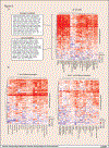
Figure 2:
Figure 2:
Expression of PD-L1 is positively correlated with expression of immunotherapy-relevant target genes across solid tumors from TCGA. (A) Heat map of Pearson R coefficients between PD-L1 expression and immune target genes by tumor type. Immune target genes were separated into those strongly correlated with PD-L1 and those less strongly correlated. (B) Heat maps of Pearson R coefficients between PD-L1 expression and immune target genes in non-T cell–inflamed tumors and T cell–inflamed tumors. Methods: Gene expression correlation analysis. Gene expression data (release date February 4, 2015) were downloaded for 30 solid tumor types from TCGA (acute myeloid leukemia, diffuse large B-cell lymphoma, and thymoma were excluded because of high tumor intrinsic immune cell transcripts). Skin cutaneous melanoma had both primary and metastatic samples available, whereas the other 29 cancers had only primary tumors available. A total of 9,555 tumor samples were included in the analysis and processed as described previously (15). Data were normalized across all samples and the patients were categorized into non-T cell–inflamed (cold), intermediate (med), and T cell–inflamed (hot) tumor groups using a previously defined 160-gene T cell–inflamed signature. A list of 166 immune molecules representative of the interactions between tumor cells and immune cells in the tumor microenvironment were selected and correlated with PD-L1 (also known as CD274). For each tumor type, Pearson product-moment correlation coefficient r were computed between the gene expression of each immune molecule and PD-L1 and used for clustering the genes by hierarchical unsupervised clustering with Euclidean distance. The genes were clustered into two distinct groups consisting of (1) strongly correlated genes such as IFNG and FOXP3, and (2) less correlated genes such as TGFB1 and VEGFA.

References

    1. Galon J, Costes A, Sanchez-Cabo F, Kirilovsky A, Mlecnik B, Lagorce-Pages C, et al. Type, density, and location of immune cells within human colorectal tumors predict clinical outcome. Science 2006;313(5795):1960–4 doi 10.1126/science.1129139. - DOI - PubMed
    1. Azimi F, Scolyer RA, Rumcheva P, Moncrieff M, Murali R, McCarthy SW, et al. Tumor-infiltrating lymphocyte grade is an independent predictor of sentinel lymph node status and survival in patients with cutaneous melanoma. Journal of clinical oncology : official journal of the American Society of Clinical Oncology 2012;30(21):2678–83 doi 10.1200/JCO.2011.37.8539. - DOI - PubMed
    1. Mahmoud SM, Paish EC, Powe DG, Macmillan RD, Grainge MJ, Lee AH, et al. Tumor-infiltrating CD8+ lymphocytes predict clinical outcome in breast cancer. Journal of clinical oncology : official journal of the American Society of Clinical Oncology 2011;29(15):1949–55 doi 10.1200/JCO.2010.30.5037. - DOI - PubMed
    1. Fuertes MB, Kacha AK, Kline J, Woo SR, Kranz DM, Murphy KM, et al. Host type I IFN signals are required for antitumor CD8+ T cell responses through CD8{alpha}+ dendritic cells. J Exp Med 2011;208(10):2005–16 doi 10.1084/jem.20101159. - DOI - PMC - PubMed
    1. Gajewski TF, Corrales L. New perspectives on type I IFNs in cancer. Cytokine & growth factor reviews 2015;26(2):175–8 doi 10.1016/j.cytogfr.2015.01.001. - DOI - PMC - PubMed

Publication types