. 2018 Dec;17(12):2387-2401.
doi: 10.1074/mcp.RA118.000892. Epub 2018 Sep 4.

Effects of Acetylation and Phosphorylation on Subunit Interactions in Three Large Eukaryotic Complexes

Affiliations

Effects of Acetylation and Phosphorylation on Subunit Interactions in Three Large Eukaryotic Complexes

Nikolina Šoštarić et al. Mol Cell Proteomics. 2018 Dec.

Abstract

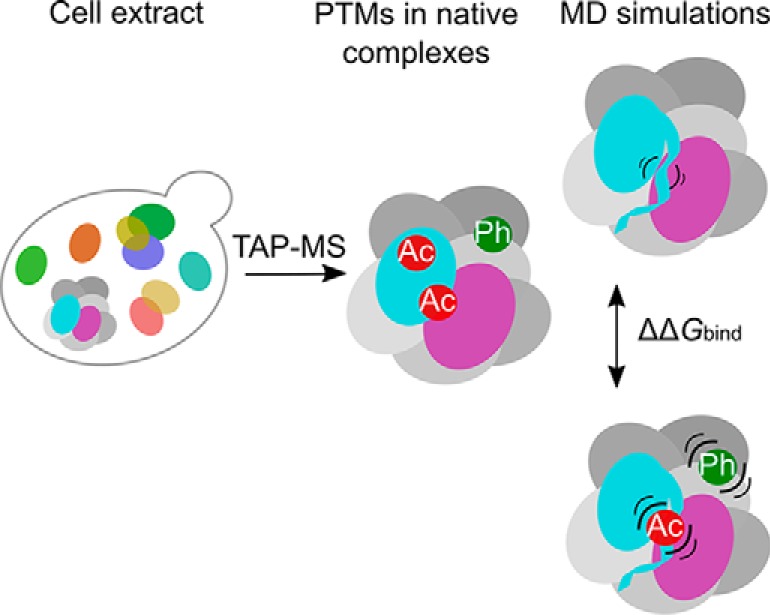
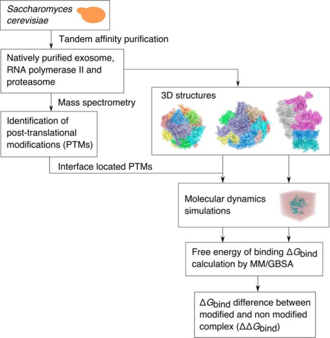
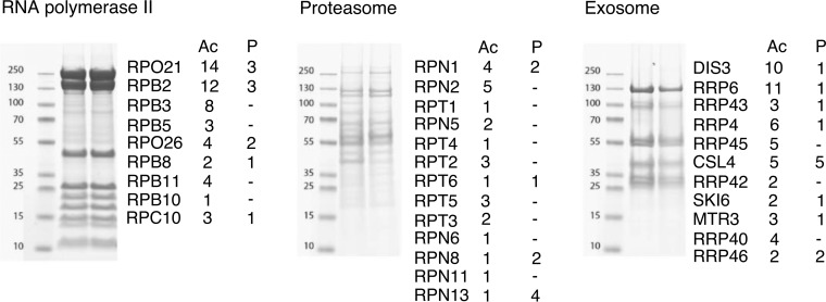
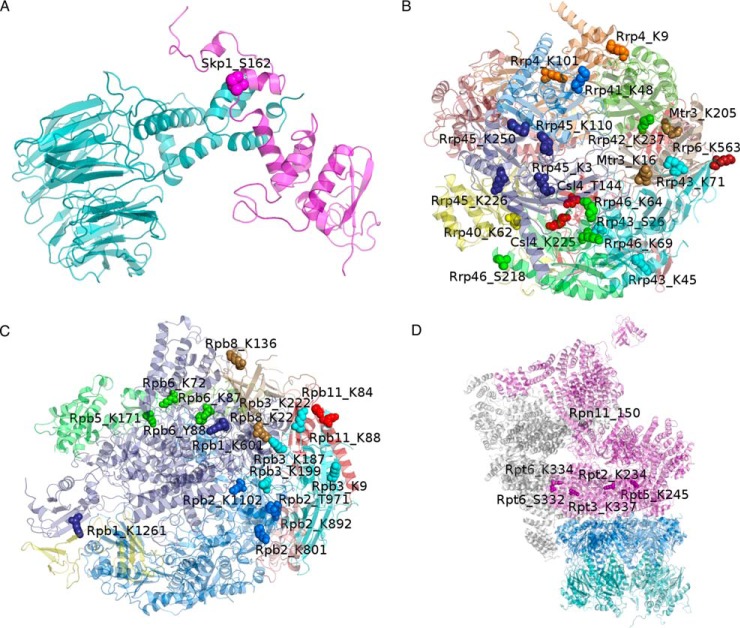
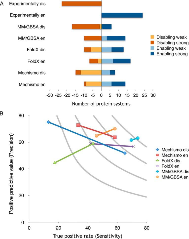
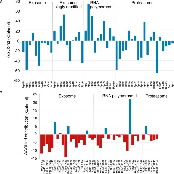
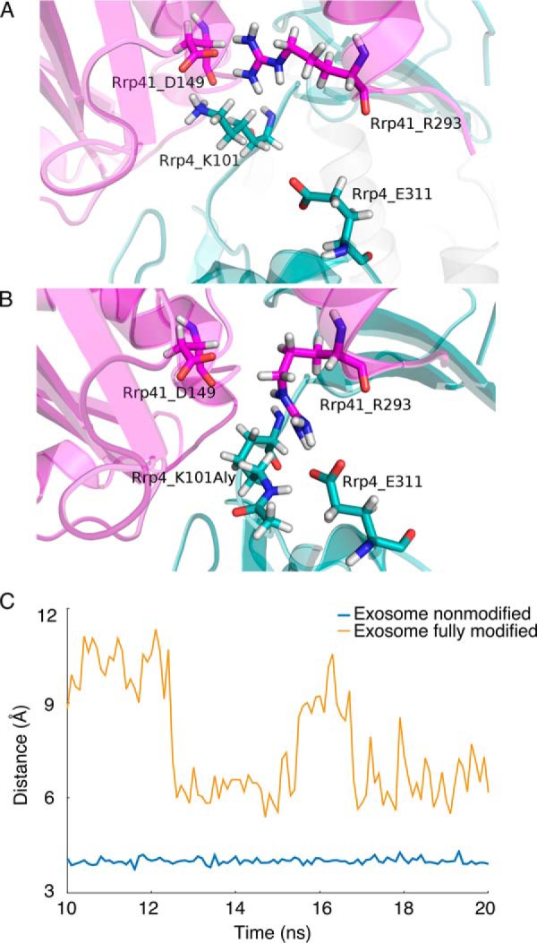
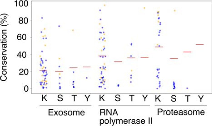
Protein post-translational modifications (PTMs) have an indispensable role in living cells as they expand chemical diversity of the proteome, providing a fine regulatory layer that can govern protein-protein interactions in changing environmental conditions. Here we investigated the effects of acetylation and phosphorylation on the stability of subunit interactions in purified Saccharomyces cerevisiae complexes, namely exosome, RNA polymerase II and proteasome. We propose a computational framework that consists of conformational sampling of the complexes by molecular dynamics simulations, followed by Gibbs energy calculation by MM/GBSA. After benchmarking against published tools such as FoldX and Mechismo, we could apply the framework for the first time on large protein assemblies with the aim of predicting the effects of PTMs located on interfaces of subunits on binding stability. We discovered that acetylation predominantly contributes to subunits' interactions in a locally stabilizing manner, while phosphorylation shows the opposite effect. Even though the local binding contributions of PTMs may be predictable to an extent, the long range effects and overall impact on subunits' binding were only captured because of our dynamical approach. Employing the developed, widely applicable workflow on other large systems will shed more light on the roles of PTMs in protein complex formation.

Keywords: Acetylation; Binding Affinity; Computational Biology; Exosome; Phosphorylation; Proteasome; Proteomics; RNA polymerase II; Structural Biology; Yeast.


Figures

None
Graphical abstract
Fig. 1.
Fig. 1.
Overview of the workflow. The strategy applied for prediction of the effect of post-translational modifications on binding of subunits in yeast exosome, RNA polymerase II and proteasome 19S regulatory particle.
Fig. 2.
Fig. 2.
Post-translational modifications in native yeast complexes. Individual subunits of the TAP-purified exosome, RNA polymerase II and proteasome separated on the gel, with number of acetylation (Ac) and phosphorylation (P) sites detected by mass spectrometry denoted on the right side from each band.
Fig. 3.
Fig. 3.
Structures of the simulated complexes. Interface located modification sites are emphasized by the space filled representation. Each site carries a label denoting the subunit, amino acid type, and its position within the respective chain. A, In Skp1:Met30 complex, Skp1 is shown in magenta and Met30 in cyan. B, Exosome and (C) RNA polymerase II subunits are each shown in different color, while (D) parts instead of individual 33 subunits of 26S proteasome are differently colored: 20S core particle β subunits in cyan and α in blue, and 19S regulatory particle base subunits in magenta and lid in gray.
Fig. 4.
Fig. 4.
Benchmarking of molecular dynamics based approach for prediction of PTMs effect on binding. A, Performance of Mechismo (Betts et al. 2017), FoldX and the MM/GBSA method presented in this work in predicting the effect of interface located phosphorylation sites on binding. Destabilizing (disabling, short dis) cases are shown in blue and stabilizing (enabling, short en) in orange, with darker shades representing predictions above the threshold for the given method. B, Precision and sensitivity of the predictions of enabling and disabling effects of interface phosphorylation sites by the three methods. Two data points for each case refer to the predicted values above/below threshold. As an ideal method has high values of both precision and sensitivity, MM/GBSA outperforms the other methods, especially in predictions of disabling effects.
Fig. 5.
Fig. 5.
Predicted effects of phosphorylations and acetylations on binding affinities in yeast exosome, RNA polymerase II and exosome. A, ΔΔGbind for all subunits of three fully modified yeast complexes and the singly modified exosome, as obtained by MM/GBSA while the respective subunit is treated as a ligand. B, Individual contributions of the interface modification sites to the overall ΔΔGbind of the respective subunit. Phosphorylation sites are depicted in blue, and acetylation in red. As in (A), negative values indicate stabilizing effect on the binding after the modifications were introduced in the structure of the complex, while the opposite is true for positive values.
Fig. 6.
Fig. 6.
Molecular dynamics revealed significant conformational changes after introduction of post-translational modifications. Changes of the interaction pattern at the interface of Rrp4 and Rrp41 exosome subunits between (A) non modified and (B) fully modified complex. Spatial re-orientation of the acetylation site Rrp4 Lys101 is accompanied by a change of interaction partner of the Rrp41 Arg293 residue from the Asp149 in its own chain, to Glu311 in the opposing subunit. Because of this change, Rrp41 Asp149 has a destabilizing effect on ΔΔGbind, while the opposite is true for Rrp41 Arg293. This can also be seen from per residue decomposition (supplemental Fig. S2C). C, Distance of salt bridge forming residues Arg210 in Rrp46 and Asp9 in Rrp40 subunit at the respective interface, measured in 100 snapshots used for MM/GBSA analysis as a distance between arginine Cζ and Asp Cγ atoms. Although these residues are distant from the modified sites in the fully modified exosome, the presence of the PTMs in the structure affected their interaction.
Fig. 7.
Fig. 7.
Conservation of modification sites identified in this study. Interface positioned residues are shown in orange, while the others are depicted in blue. Conservation with taking into account plus-minus one positions is shown for lysines. Red horizontal lines represent average conservations of non-modified residues of the same type in the investigated subunits of the respective complexes.

References

    1. Seet B. T., Dikic I., Zhou M.-M., and Pawson T. (2006) Reading protein modifications with interaction domains. Nat. Rev. Mol. Cell Biol. 7, 473–483 - PubMed
    1. Benayoun B. A., and Veitia R. A. (2009) A post-translational modification code for transcription factors: sorting through a sea of signals. Trends Cell Biol. 19, 189–197 - PubMed
    1. Kim S. C., Sprung R., Chen Y., Xu Y., Ball H., Pei J., Cheng T., Kho Y., Xiao H., Xiao L., Grishin N. V., White M., Yang X.-J., and Zhao Y. (2006) Substrate and functional diversity of lysine acetylation revealed by a proteomics survey. Mol. Cell 23, 607–618 - PubMed
    1. Cohen P. (2001) The role of protein phosphorylation in human health and disease. Eur. J. Biochem. 268, 5001–5010 - PubMed
    1. Audagnotto M., and Dal Peraro M. (2017) Protein post-translational modifications: In silico prediction tools and molecular modeling. Comput. Struct. Biotechnol. J. 15, 307–319 - PMC - PubMed

Publication types

MeSH terms

Substances