. 2018 Oct 24;62(11):e01442-18.
doi: 10.1128/AAC.01442-18. Print 2018 Nov.

Effects of PERK eIF2α Kinase Inhibitor against Toxoplasma gondii

Affiliations

Effects of PERK eIF2α Kinase Inhibitor against Toxoplasma gondii

Leonardo Augusto et al. Antimicrob Agents Chemother. .

Abstract

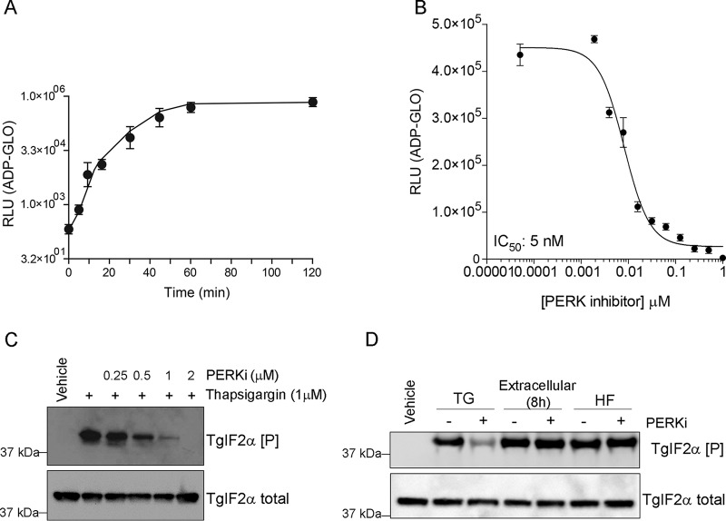
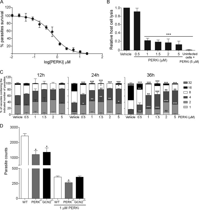
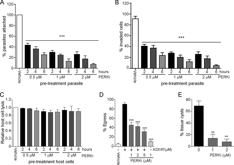
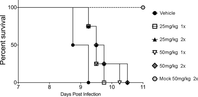
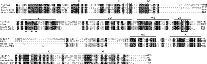
Toxoplasma gondii is an obligate intracellular parasite that has infected one-third of the population. Upon infection of warm-blooded vertebrates, the replicating form of the parasite (tachyzoite) converts into a latent form (bradyzoite) present in tissue cysts. During immune deficiency, bradyzoites can reconvert into tachyzoites and cause life-threatening toxoplasmosis. We previously reported that translational control through phosphorylation of the α subunit of T. gondii eukaryotic initiation factor 2 (eIF2α) (TgIF2α) is a critical component of the parasite stress response. Diverse stresses can induce the conversion of tachyzoites to bradyzoites, including those disrupting the parasite's endoplasmic reticulum (ER) (ER stress). Toxoplasma possesses four eIF2α kinases, one of which (TgIF2K-A) localizes to the parasite ER analogously to protein kinase R-like endoplasmic reticulum kinase (PERK), the eIF2α kinase that responds to ER stress in mammalian cells. Here, we investigated the effects of a PERK inhibitor (PERKi) on Toxoplasma Our results show that the PERKi GSK2606414 blocks the enzymatic activity of TgIF2K-A and reduces TgIF2α phosphorylation specifically in response to ER stress. PERKi also significantly impeded multiple steps of the tachyzoite lytic cycle and sharply lowered the frequency of bradyzoite differentiation in vitro Pretreatment of host cells with PERKi prior to infection did not affect parasite infectivity, and PERKi still impaired parasite replication in host cells lacking PERK. In mice, PERKi conferred modest protection from a lethal dose of Toxoplasma Our findings represent the first pharmacological evidence supporting TgIF2K-A as an attractive new target for the treatment of toxoplasmosis.

Keywords: Apicomplexa; antiparasitics; drugs; parasite; protozoa; stress response; translation.


Figures

FIG 1
FIG 1
PERKi impairs TgIF2K-A kinase activity. (A) Histidine-tagged TgIF2α was purified from E. coli and incubated with GST-TgIF2K-A-KD for the times shown. The amount of ATP consumed during the reaction time was measured using the ADP-Glo kinase assay and is expressed in relative light units (RLU). Error bars represent the standard deviation (n = 3). (B) TgIF2α was incubated with TgIF2K-A-KD for 30 min in kinase reaction buffer containing different concentrations of PERKi (0 to 2 μM). The amount of ATP consumed was measured using the ADP-Glo kinase assay, and the IC50 was calculated (IC50 = 5 nM). Error bars represent the standard deviation (n = 3). (C) Extracellular tachyzoites were treated with 1 μM thapsigargin in the presence of different concentrations of PERKi, as indicated, or incubated with the vehicle (DMSO) for 1 h. Parasite lysates were resolved by SDS-PAGE for immunoblotting with antibodies to total and phosphorylated TgIF2α. (D) Extracellular tachyzoites were treated with 1 μM thapsigargin (TG) or 50 nM halofuginone (HF) or exposed to extracellular stress for 8 h in the presence or absence of PERKi (1 μM), as indicated, and vehicle (DMSO). Samples were processed for immunoblotting as described in the legend to panel C.
FIG 2
FIG 2
PERKi impairs replication of tachyzoites in vitro. (A) A colorimetric microtiter assay was performed to assess parasite growth in a transgenic RH parasite line expressing β-galactosidase. PERKi was used over a concentration range of 0 to 20 μM and reduced the replication of tachyzoites with an IC50 of 0.62 μM and an IC90 of 2.7 μM. The viability curve shows the means for three biological replicates. The y axis shows the percent viability relative to vehicle treatment, and the x axis shows the log of the concentrations of PERKi. (B) Plaque assays for wild-type RH strain parasite cultures in the presence of various concentrations of PERKi or vehicle. The uninfected HFF monolayer was treated with 5 μM PERKi. After 5 days, the monolayers were stained to measure the area of host cell lysis. Treated samples with results that were significantly different from those for vehicle-treated samples are indicated by asterisks (***, P < 0.005). (C) Parasite counting assay. At the indicated time points, the number of parasites in 250 random vacuoles was plotted as a percentage of the total number of vacuoles examined. Significant differences between PERKi-treated and vehicle-treated cells are indicated. *, P < 0.05; **, P < 0.01; ***, P < 0.005. (D) Tachyzoites were allowed to infect MEF cells (wild-type [WT], PERK−/−, or GCN2−/− cells) in the presence or absence of 1 μM PERKi. At 48 h postinfection, parasite replication was assessed using the PCR-based assay for B1. Significant differences between the WT and knockout cells are indicated. *, P < 0.05. Error bars for all graphs show the standard deviation.
FIG 3
FIG 3
Effects of PERKi on the tachyzoite lytic cycle and differentiation. (A and B) Attachment (A) and invasion (B) assays. Extracellular RH strain tachyzoites were treated with the indicated concentrations of PERKi for 2, 4, or 6 h prior to infection of HFF host cells, and dual staining allowed determination of the percentage that attached or invaded. Significant differences between the vehicle-treated and PERKi-treated cells are indicated. ***, P < 0.005. (C) HFF monolayers were pretreated with the indicated concentrations of PERKi for 2, 4, or 6 h prior to infection. At 5 days postinfection, monolayers were stained to determine the percentage of host cell lysis. (D) Infected HFF monolayers were treated with the indicated concentrations of PERKi or vehicle for 10 min. Parasite egress was stimulated by the addition of 1 μM A23187. The percentage of egressed vacuoles in each treatment group was determined by scoring at least 100 randomly chosen vacuoles. Significant differences between the vehicle-treated and PERKi-treated cells are indicated. ***, P < 0.005. (E) The indicated concentration of PERKi (or vehicle) was included in cultures during the in vitro differentiation of ME49 type II strain parasites. At day 5, differentiated cultures were incubated with lectin stain to visualize tissue cyst walls. Significant differences between the vehicle-treated and PERKi-treated cells are indicated. **, P < 0.01. The error bars on each graph show the standard deviation.
FIG 4
FIG 4
PERKi prolongs the survival of mice with acute Toxoplasma infection. Female BALB/c mice were infected i.p. with 100 RH strain tachyzoites (or were mock infected). At 12 h postinfection, the mice were given vehicle or the indicated dose of PERKi i.p. once or twice a day (indicated by 1× or 2×, respectively). Mice (4 per group) were monitored at least three times daily, and the time to death was recorded.
FIG 5
FIG 5
Alignments of ER-resident eIF2α kinase domains. The sequences of the kinase domains of Toxoplasma gondii TgIF2K-A, Plasmodium falciparum PfPK4, mouse PERK, and human PERK were aligned using the ClustalW program. Residues highlighted in black are identical or similar among all four species, and those displaying sequence divergence among individuals of a single species are highlighted in gray. Motifs comprising the kinase domain are designated I to XI. The catalytic lysine (K) in subdomain II and the DFG motif in subdomain VII, which plays an important role in the regulation of kinase activity, are enclosed in black boxes. The methionine (M) gatekeeper residue inside subdomain V is indicated with a black arrow. Amino acid residues in the hinge are indicated with asterisks, and back-pocket regions of the active site critical for binding of PERKi are boxed in gray. The sequences of TgIF2K-A (ToxoDB accession number TGGT1_229630), PfPK4 (PlasmoDB accession number PF3D7_0628200), mouse PERK (E2AK3_MOUSE-Mus musculus, gene ID 13666), and human PERK (E2AK3_HUMAN-Homo sapiens, gene ID 9451) were obtained from the indicated databases.

References

    1. Montoya JG, Liesenfeld O. 2004. Toxoplasmosis. Lancet 363:1965–1976. doi:10.1016/S0140-6736(04)16412-X. - DOI - PubMed
    1. Hill DE, Chirukandoth S, Dubey JP. 2005. Biology and epidemiology of Toxoplasma gondii in man and animals. Anim Health Res Rev 6:41–61. doi:10.1079/AHR2005100. - DOI - PubMed
    1. Sullivan WJ Jr, Jeffers V. 2012. Mechanisms of Toxoplasma gondii persistence and latency. FEMS Microbiol Rev 36:725–733. doi:10.1111/j.1574-6976.2011.00305.x. - DOI - PMC - PubMed
    1. Luft BJ, Remington JS. 1992. Toxoplasmic encephalitis in AIDS. Clin Infect Dis 15:211–222. doi:10.1093/clinids/15.2.211. - DOI - PubMed
    1. Havelaar AH, Kemmeren JM, Kortbeek LM. 2007. Disease burden of congenital toxoplasmosis. Clin Infect Dis 44:1467–1474. doi:10.1086/517511. - DOI - PubMed

Publication types

MeSH terms

LinkOut - more resources