Review
. 2019 Oct 1;11(10):a034728.
doi: 10.1101/cshperspect.a034728.

What Are 3' UTRs Doing?

Affiliations
Review

What Are 3' UTRs Doing?

Christine Mayr. Cold Spring Harb Perspect Biol. .

Abstract

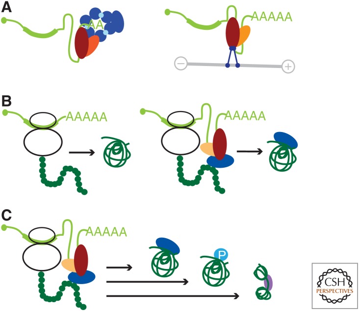
3' untranslated regions (3' UTRs) of messenger RNAs (mRNAs) are best known to regulate mRNA-based processes, such as mRNA localization, mRNA stability, and translation. In addition, 3' UTRs can establish 3' UTR-mediated protein-protein interactions (PPIs), and thus can transmit genetic information encoded in 3' UTRs to proteins. This function has been shown to regulate diverse protein features, including protein complex formation or posttranslational modifications, but is also expected to alter protein conformations. Therefore, 3' UTR-mediated information transfer can regulate protein features that are not encoded in the amino acid sequence. This review summarizes both 3' UTR functions-the regulation of mRNA and protein-based processes-and highlights how each 3' UTR function was discovered with a focus on experimental approaches used and the concepts that were learned. This review also discusses novel approaches to study 3' UTR functions in the future by taking advantage of recent advances in technology.


Figures

Figure 1.
Figure 1.
Functions of 3′ untranslated regions (UTRs). RNA-binding proteins (RBPs) bind to the 3′ UTR and recruit diverse effector proteins that determine 3′ UTR functions. (A) 3′ UTRs regulate processes at the messenger RNA (mRNA) level. RBPs (red, orange) bind to 3′ UTRs of mRNAs (light green) and recruit diverse effector proteins. The recruitment of the exosome (blue) results in mRNA destabilization (left panel), whereas the recruitment of a motor protein (blue) results in the regulation of mRNA localization using movement on a microtubule (gray line; right panel). (B) 3′ UTRs regulate protein features by mediating 3′ UTR-dependent protein–protein interactions (PPIs). Alternative 3′ UTRs can determine alternative protein functions despite encoding proteins with identical amino acid sequences. This results from 3′ UTR-dependent PPIs that are mediated only by the long 3′ UTR isoform (right panel) and not by the short 3′ UTR isoform (left panel). The RBPs that bind to the 3′ UTR, as well as the recruited effector protein, are color-coded as in A. (C) 3′ UTRs regulate diverse protein features by mediating 3′ UTR-dependent PPIs. This can result in 3′ UTR-dependent protein complex formation, 3′ UTR-dependent posttranslational modifications (P), and 3′ UTR-dependent protein folding.
Figure 2.
Figure 2.
Primary cells show a higher number and a greater magnitude of cell type–specific changes in 3′ untranslated region (3′ UTR) isoform expression. 3′-seq data are from Lianoglou et al. (2013) and were used to calculate the maximum difference in usage of alternative polyadenylation sites across seven tissues and seven cell lines. Usage is the fraction of reads mapping to a single polyadenylation site out of all the reads mapping to the 3′ UTR. Shown are the 50% of genes with most variable 3′ UTR isoform expression. Only genes that generated two 3′ UTR isoforms were included in the analysis.
Figure 3.
Figure 3.
Experimental approaches using CRISPR (clustered regularly interspaced short palindromic repeats) technology at the endogenous locus to study 3′ untranslated region (3′ UTR) functions in vivo. (A) Approach that allows phenotypes obtained for exclusive loss of the LU-generated protein compared with the total protein knockout (KO). Loss of the LU isoform is accomplished through short hairpin RNA (shRNA)-mediated knockdown (KD). At the DNA level, the arrow depicts the transcription start site. pA, Polyadenylation site; FS, frame-shift mutation. In the 3′ UTR panel, the last exon of the messenger RNA (mRNA) isoform is shown with the short 3′ UTR depicted in light green and the long 3′ UTR shown in dark green. The generated proteins are shown in blue. SU, Protein generated from the short 3′ UTR isoform; LU, protein generated from the long 3′ UTR isoform. (B) Approach that allows phenotypes obtained from the total protein KO to be compared to with exclusive expression of the protein generated from either the short (SU) or long 3′ UTR (LU) isoform. Shown as in A. The red triangles represent paired guide RNAs to delete 3′ UTR fragments. (C) Approach that allows the comparison of phenotypes obtained from the total protein KO to those generated by exclusive loss of regulatory elements in the 3′ UTR of genes that generate mRNAs with constitutive 3′ UTRs. Shown as in B. The regulation of mRNA abundance through the endogenous polyadenylation signal is preserved.

References

    1. An JJ, Gharami K, Liao GY, Woo NH, Lau AG, Vanevski F, Torre ER, Jones KR, Feng Y, Lu B, et al. 2008. Distinct role of long 3′ UTR BDNF mRNA in spine morphology and synaptic plasticity in hippocampal neurons. Cell 134: 175–187. - PMC - PubMed
    1. Andressoo JO, Mitchell JR, de Wit J, Hoogstraten D, Volker M, Toussaint W, Speksnijder E, Beems RB, van Steeg H, Jans J, et al. 2006. An Xpd mouse model for the combined xeroderma pigmentosum/Cockayne syndrome exhibiting both cancer predisposition and segmental progeria. Cancer Cell 10: 121–132. - PubMed
    1. Baltz AG, Munschauer M, Schwanhausser B, Vasile A, Murakawa Y, Schueler M, Youngs N, Penfold-Brown D, Drew K, Milek M, et al. 2012. The mRNA-bound proteome and its global occupancy profile on protein-coding transcripts. Mol Cell 46: 674–690. - PubMed
    1. Barreau C, Paillard L, Osborne HB. 2005. AU-rich elements and associated factors: Are there unifying principles? Nucleic Acids Res 33: 7138–7150. - PMC - PubMed
    1. Batra R, Charizanis K, Manchanda M, Mohan A, Li M, Finn DJ, Goodwin M, Zhang C, Sobczak K, Thornton CA, et al. 2014. Loss of MBNL leads to disruption of developmentally regulated alternative polyadenylation in RNA-mediated disease. Mol Cell 56: 311–322. - PMC - PubMed

Publication types