. 2018 Sep 4;8(1):13181.
doi: 10.1038/s41598-018-31342-4.

Identification of a dioxin-responsive oxylipin signature in roots of date palm: involvement of a 9-hydroperoxide fatty acid reductase, caleosin/peroxygenase PdPXG2

Affiliations

Identification of a dioxin-responsive oxylipin signature in roots of date palm: involvement of a 9-hydroperoxide fatty acid reductase, caleosin/peroxygenase PdPXG2

Abdulsamie Hanano et al. Sci Rep. .

Abstract

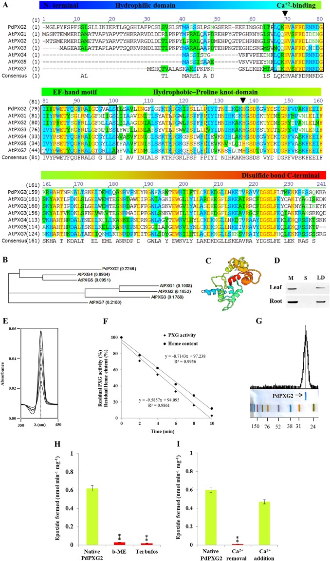
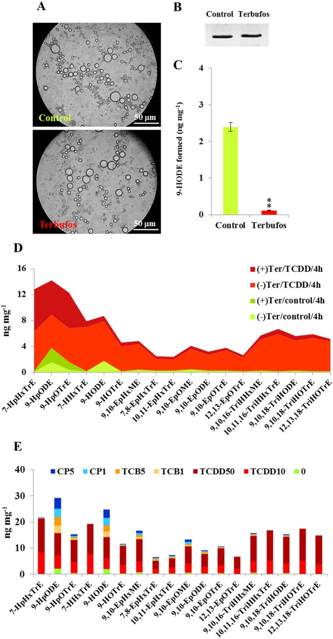
Dioxins are highly hazardous pollutants that have well characterized impacts on both animal and human health. However, the biological effects of dioxins on plants have yet to be described in detail. Here we describe a dioxin-inducible caleosin/peroxygenase isoform, PdPXG2, that is mainly expressed in the apical zone of date palm roots and specifically reduces 9-hydroperoxide fatty acids. A characteristic spectrum of 18 dioxin-responsive oxylipin (DROXYL) congeners was also detected in date palm roots after exposure to dioxin. Of particular interest, six oxylipins, mostly hydroxy fatty acids, were exclusively formed in response to TCDD. The DROXYL signature was evaluated in planta and validated in vitro using a specific inhibitor of PdPXG2 in a root-protoplast system. Comparative analysis of root suberin showed that levels of certain monomers, especially the mono-epoxides and tri-hydroxides of C16:3 and C18:3, were significantly increased after exposure to TCDD. Specific inhibition of PdPXG2 activity revealed a positive linear relationship between deposition of suberin in roots and their permeability to TCDD. The results highlight the involvement of this peroxygenase in the plant response to dioxin and suggest the use of dioxin-responsive oxylipin signatures as biomarkers for plant exposure to this important class of xenobiotic contaminants.


Conflict of interest statement

The authors declare no competing interests.

Figures

Figure 1
Figure 1
Sequence analysis and biochemcal properities of PdPXG2. (A) Multiple alignment of the PdPXG2 sequence with A. thaliana AtPXG1 (At4g26740), AtPXG2 (At5g55240), AtPXG3 (At2g33380), AtPXG4 (At1g70670), AtPXG5 (At1g70680) and AtPXG7 (At1g23240). The boxed areas correspond to the Ca2+ binding and the proline knot domains. Triangles indicate positions of Histidines H68 and H136 responsible to heme binding. (B) Phylogenetic tree of PdPXG2 with its orthologs in A. thaliana. (C) Predicted 3D protein structure of PdPXG2. The virtual image was generated online using http://www.sbg.bio.ic.ac.uk/phyre2. Image was rainbow colored from N to C terminus. (D) Immunoblotting of PdPXG2 protein by a polyclonal antibody prepared from the complete sequence of AtCLO1 caleosin from Arabidopsis thaliana, diluted 1:500 in TBS buffer (pH 7.4). (E) Sequential scanning of the heme-spectrum of PdPXG2 obtained by addition of cumene hydroperoxide at the indicated times. (F) Correlation between the epoxidation activity of purified PdPXG2 and disappearance of the heme content. (G) PdPXG2 was radio-phosphorylated by casein kinase and [35S]ATP. (H). Inhibition of native PdPXG2 was by treatment with β-mercaptoethanol (β-MPE) (1 mM) or terbufos (3 mM). (I) Activities for native purified PdPXG2 and Ca2+-deionized PdPXG2 after extensive dialysis and for PdPXG2 after dialysis followed by addition of 1 mM CaCl2 to the medium. All measurements were in triplicate. Values are the means ± S.D. (n = 3). Asterisks indicate significant differences in epoxidation activities between treated and native PdPXG2 (**P < 0.01).
Figure 2
Figure 2
Catalytic properities of PdPXG2. (A) Fatty acid epoxidation activity of PdPXG2 as a function of carbon chain length and unsaturation degree of fatty acids. (B) Stereoselectivity of PdPXG2 towards the cis-double bond configuration in C16:1 or C18:1. PdPXG2 exhibited a strong stereoselectivity since no epoxidation was detected with UFAs having the double bond in trans-configuration regardless of the position (6, 9 or 11) of the cis-double bond in the carbon chain. (C) Epoxidation of [14C]-labelled palmitic acid (C16:1) and oleic acid (C18:1) (Cis-Δ9 for both) by recombinant PdPXG2 in the presence of various hydroperoxides; i.e. H2O2, 7-HpHxTrE, 11-HpHxTrE, 9-HpODE, 13-HpODE, 9-HpOTrE and 13-HpOTrE compared with cumene hydroperoxide (Cu-OOH). (D) The reductase activity of purified recombinant PdPXG2 against fatty acid hydroperoxides, 7-HpHxTrE, 11-HpHxTrE, 9-HpODE, 13-HpODE, 9-HpOTrE and 13-HpOTrE using a HPLC-UV-detector system. All measurements were in triplicate. Values are the means ± S.D. (n = 3). *P < 0.05; **P < 0.01.
Figure 3
Figure 3
Expression and activity of PdPXG2 as a function of root development. (A) Quantification of PdPXG2 transcripts in whole roots of date palm seedlings at different stages of development (from I to V) compared with non-germinated seeds (stage 0). For each stage, the transcript level was evaluated by qRT-PCR. (B) Spatiotemporal variations of PdPXG2 gene expression ordered in the sections taken from the top to the root apical zones. (C) Immunoblotting of PdPXG2 protein was performed in the same samples used in B using a polyclonal antibody prepared from the complete sequence of AtCLO1 caleosin from Arabidopsis thaliana, diluted 1:500 in. (D) The enzymatic activity, measured as a 9-HpODE-reductase, followed the accumulation of PdPXG2 proteins in the extracts of whole roots at the indicated stages of development (I to V). (E) Spatiotemporal variations of 9-HpODE-reductase activity as a function of localization from the top to the root apical at stages (II, III, IV and V). (F) Image of a date palm seedling at stage V. The triangle shows the spatiotemporal variations of PdPXG2 activity along roots from the apical zone (dark-green) to the top (light-green). All measurements were in triplicate. Values are the means ± S.D. (n = 3). *P < 0.05; **P < 0.01; ***P < 0.001.
Figure 4
Figure 4
Expression and activity of PdPXG2 in the root of date palm seedling after exposure to TCDD. (A) Relative expression of PdPXG2 in different sections (S1–S5) of roots at various developmental stages (I to V) after exposure to TCDD (0, 10 and 50 ng.L−1, light-green, light-red and dark-red, respectively). For each section, the transcript level was evaluated by qRT-PCR. Three measurements were taken in three cDNAs prepared from three individual plants for each treatment. The colour scale (white-green-black) indicates relative changes of transcript abundance of 1, 50 and 100 fold, respectively. The expression level in control samples was defined as 1, and corresponding abundance changes under 10 and 50 ng.L−1 TCDD were calculated directly using the Applied Biosystems qPCR system software. (B) Immunoblotting of PdPXG2 protein in the same samples of control and those treated with 50 ng.L−1 using a polyclonal antibody of AtCLO1 caleosin. (C) Spatiotemporal evaluation of 9-HpODE reductase activity as a function of section localization from the top to the root apical at stages (II, III, IV and V) after exposure to 10 and 50 ng.L−1 (light-red and dark -red, respectively) compared with controls (light-green). Activities were measured in triplicate. Values are the means ± S.D. (n = 3). Asterisks indicate significant differences in the tissue reductase-activity between TCDD-treatments and controls (*P < 0.05; **P < 0.01; ***P < 0.001). Lower cases indicate significant differences in the sectional reductase-activity between TCDD-treatments and controls (aP < 0.05; bP < 0.01; cP < 0.001).
Figure 5
Figure 5
Quantification of PdPXG2-derivatives oxylipins in the date palm root after exposure to TCDD. (A) Amounts of fatty acid hydroperoxides (-OOH), expressed as ng mg−1 FW (fresh weight), resulting from oxygenation of C16:3, C18:2 and C18:3 under the action of 9-LOX, 13-LOX or α-DIOX in roots of date palm after exposure to TCDD at 10 ng.L−1 (light-red columns) or 50 ng.L−1 (dark -red columns) compared with control (light-green columns). (B) Amounts of fatty acid hydroxides (-OH) resulting from reduction of the indicated hydroperoxides in root tissues after exposure to TCDD compared with controls. (C) Amounts of mono- and di-epoxy fatty acids formed from C14:1, C16:1, C16:3, C18:1, C18:2 and C18:3 in root tissues in response to TCDD-exposure. (D) Amounts of di- and tri-hydroxy of C16:1, C16:3, C18:1, C18:2 and C18:3 in the same samples. (E) Overall evaluation of the quantitative areas covered by TCDD-induced oxylipin congeners after administration of a high (dark -red) or a low dose (light-red) of TCDD. All measurements were done in triplicate. Values are the means ± S.D. (n = 3). *P < 0.05; **P < 0.01; ***P < 0.001; ****P < 0.0001.
Figure 6
Figure 6
Induction of PdPXG2-derivatives oxylipins in the date palm root-protoplasts after exposure to TCDD. (A) Image of a stage I-date palm seedling showed the root that was taken to prepare protoplasts. (B) Micrograph of prepared protoplasts. Bar represents 50 μm. (C) Immunoblotting of PdPXG2 protein in control protoplasts and protoplasts after exposure to 10 ng L−1 TCDD for 2, 4 and 8 h. (D) 9-HpODE-reductase activity of PdPXG2 in control protoplasts and TCDD-treated protoplasts for 2, 4 and 8 h. (E) Overall evaluation of quantitative areas covered by TCDD-induced oxylipins (18 congeners) after addition of TCDD (10 ng L−1) (light-red for 2 h), (red for 4 h) and (dark-red for 8 h), compared with controls (light-green for 0 h). All measurements were done in triplicate. Values are the means ± S.D. (n = 3). Asterisks indicate significant differences in the reductase-activity between TCDD-treated and control protoplasts (*P < 0.05; **P < 0.01).
Figure 7
Figure 7
Inhibition of PdPXG2 abolished the induction of TCDD-responsive oxylipins in the root-protoplasts. (A) Micrographs of control protoplasts (upper) and protoplasts pre-treated with 1 mM terbufos (Ter) (lower) for 2 h at room temperature. Bar presents 50 μm. (B) Immunoblotting of PdPXG2 protein in control protoplasts and in terbufos-pretreated protoplasts. (C) 9-HpODE-redyctase activity in control and terbufos-pretreated protoplasts. (D) Overall evaluation of the quantitative areas covered by TCDD-responsive oxylipins (18 congeners) in control and in terbufos-pretreated protoplasts after addition of TCDD (10 ng L−1) for 4 h. Color coding: light-green for control protoplasts, green for control protoplasts with terbufos, light-red for TCDD-exposed protoplasts without Ter and dark-red for TCDD-exposed protoplasts with Ter. (E) Evaluation of the TCDD-specificity of the DROXYL signature in comparison with other types of stressor agents. Similar to what we did for TCDD, the oxylipins signature was determined in the root that treated with 3,3′,4,4′-Tetrachlorobiphenyl (TCB) or with chlorpyrifos-methyl (CP) at two different concentration of each (1 and 5 μg L−1 for TCB) and (1 and 5 mg L−1 for CP). All measurements were done in triplicate. Values are means ± S.D. (n = 3). Asterisks indicate significant differences in the reductase-activity between tebufos-pretreated and control protoplasts (**P < 0.01).
Figure 8
Figure 8
PdPXG2 activity is likely necessary to the deposition of suberin and therefore to their permeability toward dioxin. (AC) Micrographs of freehand sections of control date palm roots and roots exposed to TCDD with 10 and 50 ng L−1, respectively, under fluorescent microscopy. Bar presents 300 μm. (D) Amount of total suberin extracted from TCDD-exposed root at both concentrations compared with control roots. (E) Qualitative and quantitative determinations of suberin composition in roots exposed to a low (light-red) and a high dose (dark-red) of TCDD, compared with control roots (light-green). (F) Micrographs of freehand sections of control roots without (left) or with Ter (right) and exposed roots with or without Teb. (G) Micrographs of freehand sections of TCDD-exposed roots without (left) or with Ter (right). (H) Total amount of suberin in control roots without or with Teb (light-green and green columns, respectively) and in TCDD-exposed roots without or with Ter-pretreatment (red and dark-red columns, respectively). (I) Composition of suberin in control roots without or with Teb (light-green and green columns, respectively) and in TCDD-exposed roots without or with Ter-pretreatment (red and dark-red columns, respectively). (J) Progressive inhibition of PdPXG2 by increasing concentration of Terbufos in correlation with the total amount of root suberin and their permeability against the TCDD. Bar presents 300 μm. All measurements were in triplicate. Data are means ± S.D. (n = 3). *P < 0.05; **P < 0.01; ***P < 0.001; ****P < 0.0001.

References

    1. WHO. Dioxins and their effects on human health. http://www.who.int/mediacentre/factsheets/fs225/en/, Updated October 2016 (2016).
    1. Tuppurainen K, Asikainen A, Ruokojarvi P, Ruuskanen J. Perspectives on the formation of polychlorinated dibenzo-p-dioxins and dibenzofurans during municipal solid waste (MSW) incineration and other combustion processes. Acc Chem Res. 2003;36:652–658. doi: 10.1021/ar020104+. - DOI - PubMed
    1. Meharg AA, Osborn D. Dioxins released from chemical accidents. Nature. 1995;375:353–354. doi: 10.1038/375353a0. - DOI - PubMed
    1. Pollitt F. Polychlorinated dibenzodioxins and polychlorinated dibenzofurans. Regul Toxicol Pharmacol. 1999;30:S63–68. doi: 10.1006/rtph.1999.1328. - DOI - PubMed
    1. Poland A, Knutson JC. 2,3,7,8-tetrachlorodibenzo-p-dioxin and related halogenated aromatic hydrocarbons: examination of the mechanism of toxicity. Annual Review of Pharmacology and Toxicology. 1982;22:517–554. doi: 10.1146/annurev.pa.22.040182.002505. - DOI - PubMed

MeSH terms

LinkOut - more resources