. 2018 Sep;22(5):555-566.
doi: 10.4196/kjpp.2018.22.5.555. Epub 2018 Aug 27.

Human umbilical cord blood mesenchymal stem cells engineered to overexpress growth factors accelerate outcomes in hair growth

Affiliations

Human umbilical cord blood mesenchymal stem cells engineered to overexpress growth factors accelerate outcomes in hair growth

Dong Ho Bak et al. Korean J Physiol Pharmacol. 2018 Sep.

Abstract

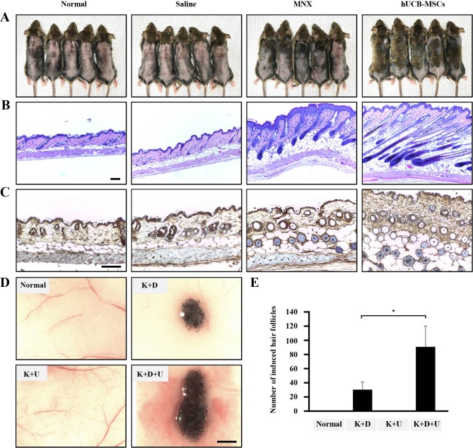
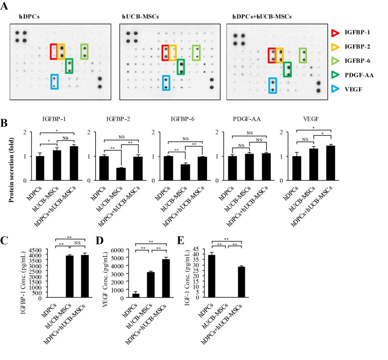
Human umbilical cord blood mesenchymal stem cells (hUCB-MSCs) are used in tissue repair and regeneration; however, the mechanisms involved are not well understood. We investigated the hair growth-promoting effects of hUCB-MSCs treatment to determine whether hUCB-MSCs enhance the promotion of hair growth. Furthermore, we attempted to identify the factors responsible for hair growth. The effects of hUCB-MSCs on hair growth were investigated in vivo, and hUCB-MSCs advanced anagen onset and hair follicle neogeneration. We found that hUCB-MSCs co-culture increased the viability and up-regulated hair induction-related proteins of human dermal papilla cells (hDPCs) in vitro. A growth factor antibody array revealed that secretory factors from hUCB-MSCs are related to hair growth. Insulin-like growth factor binding protein-1 (IGFBP-1) and vascular endothelial growth factor (VEGF) were increased in co-culture medium. Finally, we found that IGFBP-1, through the co-localization of an IGF-1 and IGFBP-1, had positive effects on cell viability; VEGF secretion; expression of alkaline phosphatase (ALP), CD133, and β-catenin; and formation of hDPCs 3D spheroids. Taken together, these data suggest that hUCB-MSCs promote hair growth via a paracrine mechanism.

Keywords: Alopecia; Dermal papilla cell; Hair growth; IGFBP-1; Stem cell; Stem-cell therapy.


Conflict of interest statement

CONFLICTS OF INTEREST: The authors declare no conflicts of interest.

Figures

Fig. 1
Fig. 1. The effects of hUCB-MSCs on induction of the hair cycle anagen stage in mice.
The dorsal skin of six-week-old male C3H/HeJ mice was shaved and then injected with saline, treated with topical application of 3% minoxidil (MNX, 5 times per week), or injected with hUCB-MSCs (1×10⁵ cells/head) (A) Gross images of hair regrowth in C3H/HeJ mice treated with either saline, MNX, or hUCB-MSCs at six weeks after shaving. (B) Histological analysis of dorsal skin samples from each group using H&E staining. Scale bar, 100 µm (C) Immunohistochemistry, dorsal skin samples from each group were stained with anti-β-catenin, Scale bar, 100 µm. (D) Patch assay: at 2 weeks, the nude mice were sacrificed, and newly generated hair follicles were counted. Scale bar, 1 mm. (E) Bar graph showing the number of each hair follicles as mean±SD. *p<0.05 vs. K plus D group. **p<0.01, ***p<0.001 vs. hDPCs alone.
Fig. 2
Fig. 2. The effects of hUCB-MSCs on cell viability, ALP activity, and AKT/GSK3β/β-catenin pathway regulation of hDPCs.
hDPCs were co-cultured with hUCB-MSCs for 48 h or 120 h in two types of culture media (DMEM and MEM). (A) The cell viability was estimated using a WST-8 assay. (B) ALP activity. (C) Representative images of Western blot protein assays for p-AKT, AKT, p-GSK3β, GSK3β, β-catenin, and proliferating cell nuclear antigen (PCNA) in dermal papilla cells. hDPCs and hUCB-MSCs were seeded in 6-well transwell plates. After 48 h, the cell lysates were harvested for Western blot assays. (D) Intensities of the immunoreactive bands on the Western blots as quantified by densitometric analysis. For all graphs, the data is reported as mean±SD. *p<0.01, ***p<0.001 vs. hDPCs alone.
Fig. 3
Fig. 3. Growth factor secretion profiles of hDPCs alone, hUCB-MSCs alone, and hDPCs plus hUCB-MSCs.
(A) Representative images of the growth factor antibody array analysis in culture media obtained at 48 h of growth in each group. (B) Quantification of the cytokine array analysis of the culture media as the fold increase compared with hDPCs alone. The protein concentration in each culture media measured individual protein with ELISA. (C) IGFBP-1, (D) VEGF, and (E) IGF-1. Abbreviations are as follows: IGFBP-1, -2, and -6, insulin-like growth factor binding protein-1, -2, and -6; PDGF-AA, platelet-derived growth Factor-AA; VEGF, vascular endothelial growth factor; IGF-1, insulin-like growth factor-1. The data are reported as mean±SD. *p<0.05, **p<0.01, vs. hDPCs alone.
Fig. 4
Fig. 4. The effects of recombinant IGFBP-1 on hDPC viability, VEGF secretion, expression of proteins related to hair anagen induction, and formation of 3D tissue-like structures of hDPCs.
(A) Cell viability. Control hDPCs (Cont.) and IGFBP-1 (12.5–200 ng/ml)-treated hDPCs were cultured for 48 h, and the cell viability was measured using an WST-8 assay. (B) VEGF concentration in the culture media. Cont. and IGFBP-1 (12.5–200 ng/ml)-treated hDPCs were cultured for 48 h. The culture media was harvested, and VEGF concentration was measured using an ELISA assay. (C) Immunofluorescent staining of protein ALP, CD133, and β-catenin in hDPCs. rhIGFBP-1 (100 ng/ml) was administered to hDPCs (1×10⁵) for 48 h. The cells were then stained to visualize the expression of anti-ALP, -CD133, and -β-catenin (Green). Scale bar 5 µm. (D) Representative images of Western blot. Cell lysates of hDPCs without or with rhIGFBP-1 (100 ng/mL) were immunoprobed with anti-ALP, -CD133, non-p-β-catenin, and β-catenin. Effect of rhIGFBP-1 (100 ng/mL) on the formation of hDPC spheroids after seeding for 5 days. (E) Representative images of passage 8-hDPCs from each treatment group (n=4) aggregated into microtissues in a regular manner (0.25×10⁴ cells/well, scale bar 200 µm). (F) The bar graph shows the maximum tissue diameter produced in each group at Day 5. For all graphs, the data is shown as mean±SD. **p<0.01, ***p<0.001, vs. Cont. #p<0.01 vs. IGFBP (−).
Fig. 5
Fig. 5. The effects of recombinant IGFBP-1 on the expression of IGF-1 on hDPCs via the co-localization of an IGF-1 and IGFBP-1.
hDPCs (1×10⁵ cells) were treated with rhIGFBP-1 (100 ng/mL) for 48 h and then stained with anti-IGF-1 (green), anti-IGFBP-1 (red), and DAPI (nucleus, blue). Scale bar 20 µm.

Similar articles

Cited by

References

    1. Ferriman D, Gallwey JD. Clinical assessment of body hair growth in women. J Clin Endocrinol Metab. 1961;21:1440–1447. - PubMed
    1. Harada T, Izaki S, Tsutsumi H, Kobayashi M, Kitamura K. Apoptosis of hair follicle cells in the second-degree burn wound unders hypernatremic conditions. Burns. 1998;24:464–469. - PubMed
    1. Leyden J, Dunlap F, Miller B, Winters P, Lebwohl M, Hecker D, Kraus S, Baldwin H, Shalita A, Draelos Z, Markou M, Thiboutot D, Rapaport M, Kang S, Kelly T, Pariser D, Webster G, Hordinsky M, Rietschel R, Katz HI, Terranella L, Best S, Round E, Waldstreicher J. Finasteride in the treatment of men with frontal male pattern hair loss. J Am Acad Dermatol. 1999;40:930–937. - PubMed
    1. Sinclair R. Male pattern androgenetic alopecia. BMJ. 1998;317:865–869. - PMC - PubMed
    1. Kligman AM. Pathologic dynamics of human hair loss. I. Telogen effuvium. Arch Dermatol. 1961;83:175–198. - PubMed