

Ischemia-reperfusion injury in stroke: impact of the brain barriers and brain immune privilege on neutrophil function
- PMID: 30181779
- PMCID: PMC6111395
- DOI: 10.1177/1756286418794184
Ischemia-reperfusion injury in stroke: impact of the brain barriers and brain immune privilege on neutrophil function
Abstract
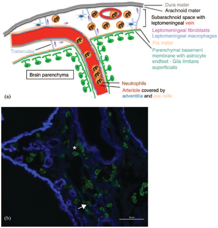
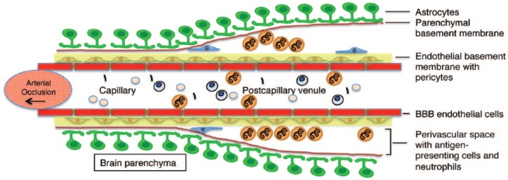
Reperfusion injury following ischemic stroke is a complex pathophysiological process involving numerous mechanisms ranging from the release of excitatory amino acids and ion disequilibrium to the induction of apoptosis and necrosis, to oxidative stress and inflammation. The migration of neutrophils into the brain parenchyma and release of their abundant proteases are generally considered the main cause of neuronal cell death and acute reperfusion injury following ischemic stroke. Recent findings in experimental and human stroke have challenged this view, as the majority of neutrophils were rather found to accumulate within the neurovascular unit (NVU) and the subarachnoid space (SAS) where they remain separated from the brain parenchyma by the glia limitans. The brain parenchyma is an immune-privileged site that is not readily accessible to immune cells and does not elicit stereotypic adaptive or innate immune responses. Understanding brain immune privilege requires intimate knowledge of its unique anatomy in which the brain barriers, that include the glia limitans, establish compartments that differ remarkably with regard to their accessibility to the immune system. We here propose that the brain immune privilege also extends to an ischemic insult, where the brain parenchyma does not evoke a rapid infiltration of neutrophils as observed in ischemic events in peripheral organs. Rather, neutrophil accumulation in the NVU and SAS could have a potential impact on cerebrospinal fluid (CSF) drainage from the central nervous system (CNS) and thus on edema formation and reperfusion injury after ischemic stroke. Integrating the anatomical and functional implications of the brain immune privilege with the unquestionable role of neutrophils in reperfusion injury is a prerequisite to exploit appropriate strategies for therapeutic interventions aiming to reduce neuronal cell death after ischemic stroke.
Keywords: brain barriers; innate immunity; ischemia; reperfusion; vascular biology.
Conflict of interest statement
Conflict of interest statement: The authors declare that there is no conflict of interest.
Figures
References
-
- Billingham RE, Brent L, Medawar PB. Actively acquired tolerance of foreign cells. Nature 1953; 172: 603–606. - PubMed
-
- Galea I, Bechmann I, Perry VH. What is immune privilege (not)? Trends Immunol 2007; 28: 12–18. - PubMed
-
- Andersson PB, Perry VH, Gordon S. The acute inflammatory response to lipopolysaccharide in CNS parenchyma differs from that in other body tissues. Neuroscience 1992; 48: 169–186. - PubMed
-
- Locatelli G, Wortge S, Buch T, et al. Primary oligodendrocyte death does not elicit anti-CNS immunity. Nat Neurosci 2012; 15: 543–550. - PubMed
Publication types
LinkOut - more resources
Full Text Sources
Other Literature Sources
