

Ultrasonographic assessment of paediatric ocular emergencies: A tertiary eye hospital based observation
- PMID: 30181795
- PMCID: PMC6117533
- DOI: 10.5847/wjem.j.1920-8642.2018.04.006
Ultrasonographic assessment of paediatric ocular emergencies: A tertiary eye hospital based observation
Abstract
Background: The purpose of this study is to assess the utility of ocular ultrasound B scan in the emergency at the first point of care for detecting posterior segment and orbital pathologies in cases of paediatric ocular emergencies.
Methods: A prospective observational study involving 122 paediatric patients presenting to eye emergency over a period of ninety days were assessed with ultrasonography for the posterior segment as well as orbital pathology whenever indicated. The ocular ultrasound was performed gently over closed eyelids.
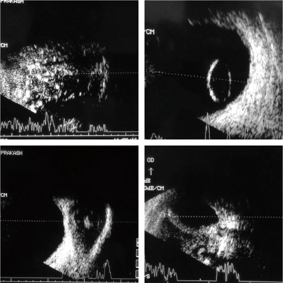
Results: Posttraumatic globe injuries were the most common indication for posterior segment evaluation, which constituted 80 (65.57%) eyes. Among these 52 patients had an anechoic posterior segment and 28 patients had variable findings such as vitreous haemorrhage (8.19%), retinal detachment (6.55%), choroidal detachment (4.91%), posteriorly dislocated clear lens (0.81%) and retained intraocular foreign body (5.73%). Non-traumatic cases constituted around 42 (34.42%) eyes, which included corneal ulcer (7.37%), retinoblastoma (6.55%), endophthalmitis (4.91%), extra-ocular muscle cysticercosis (4.91%), orbital cellulitis (4.09%), periocular haemorrhage (2.45%), proptosis(1.63%), paediatric cataract (1.63%) and cryptophthalmos (0.81%). No adverse events of performing the ultrasound was noted.
Conclusion: First point ultrasonography in paediatric ocular emergencies is a cheap, portable and an effective tool in the assertion of significant posterior segment and orbital diseases.
Keywords: Paediatric ocular emergencies; Posterior segment and orbital pathologies; Ultrasound B scan.
Conflict of interest statement
Conflicts of interest: The authors declare that there are no conflicts of interest regarding the publication of this paper.
Figures

References
-
- Moore CL, Copel JA. Point-of-care ultrasonography. N Engl J Med. 2011;364(8):749–57. - PubMed
-
- Horowitz R, Bailitz J. Ocular ultrasound—point of care imaging of the eye. Clin Pediatr Emerg Med. 2015;16(4):262–8.
LinkOut - more resources
Full Text Sources
Other Literature Sources