Review
. 2018 Sep;23(9):1-20.
doi: 10.1117/1.JBO.23.9.091415.

Review of clinical approaches in fluorescence lifetime imaging ophthalmoscopy

Affiliations
Review

Review of clinical approaches in fluorescence lifetime imaging ophthalmoscopy

Lydia Sauer et al. J Biomed Opt. 2018 Sep.

Erratum in

Abstract

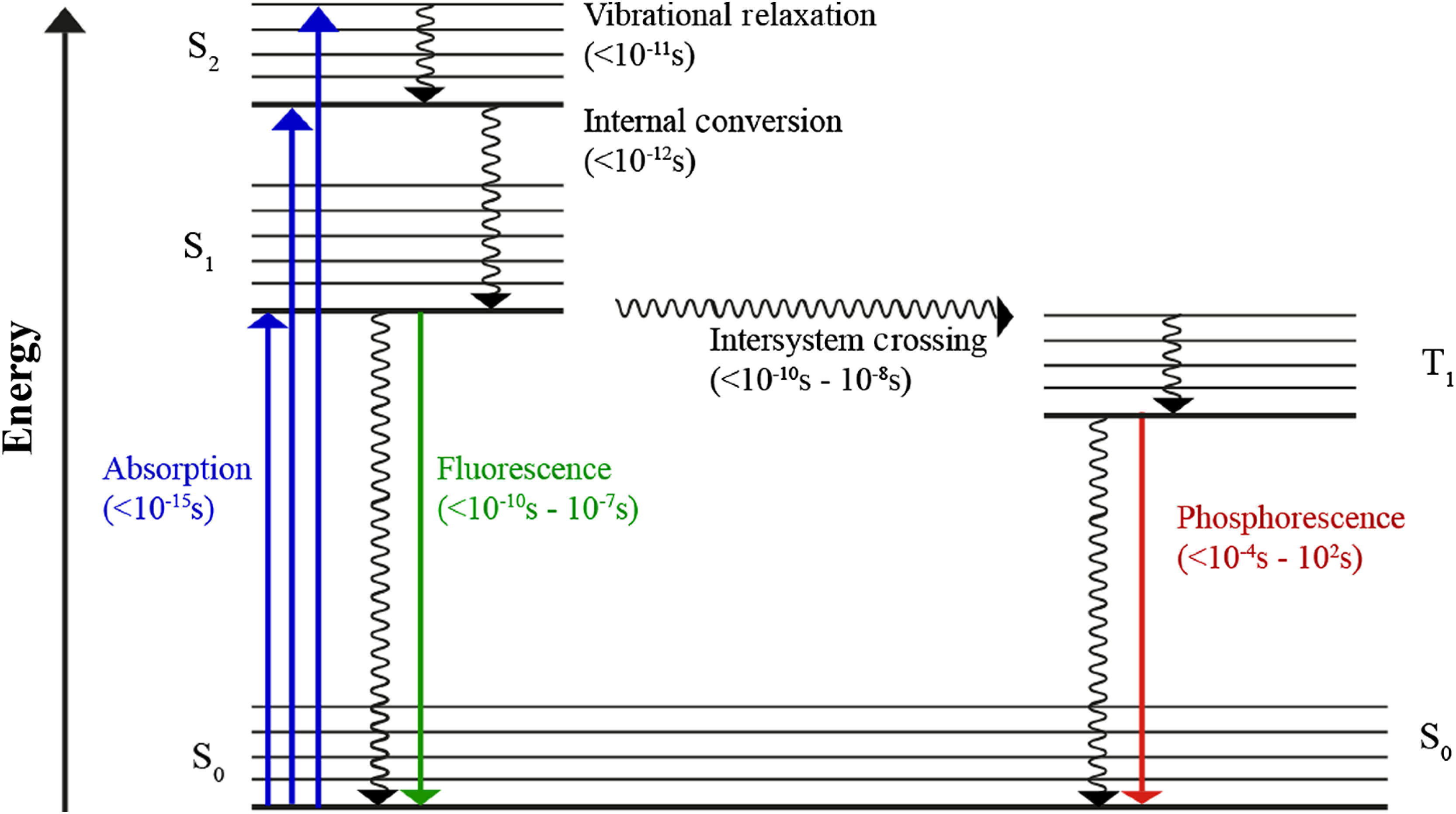
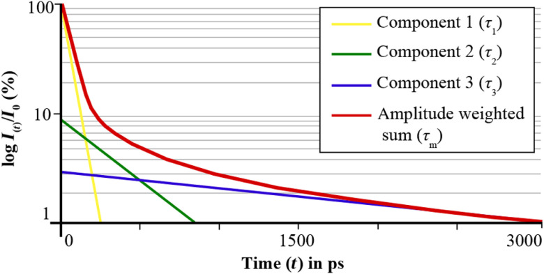
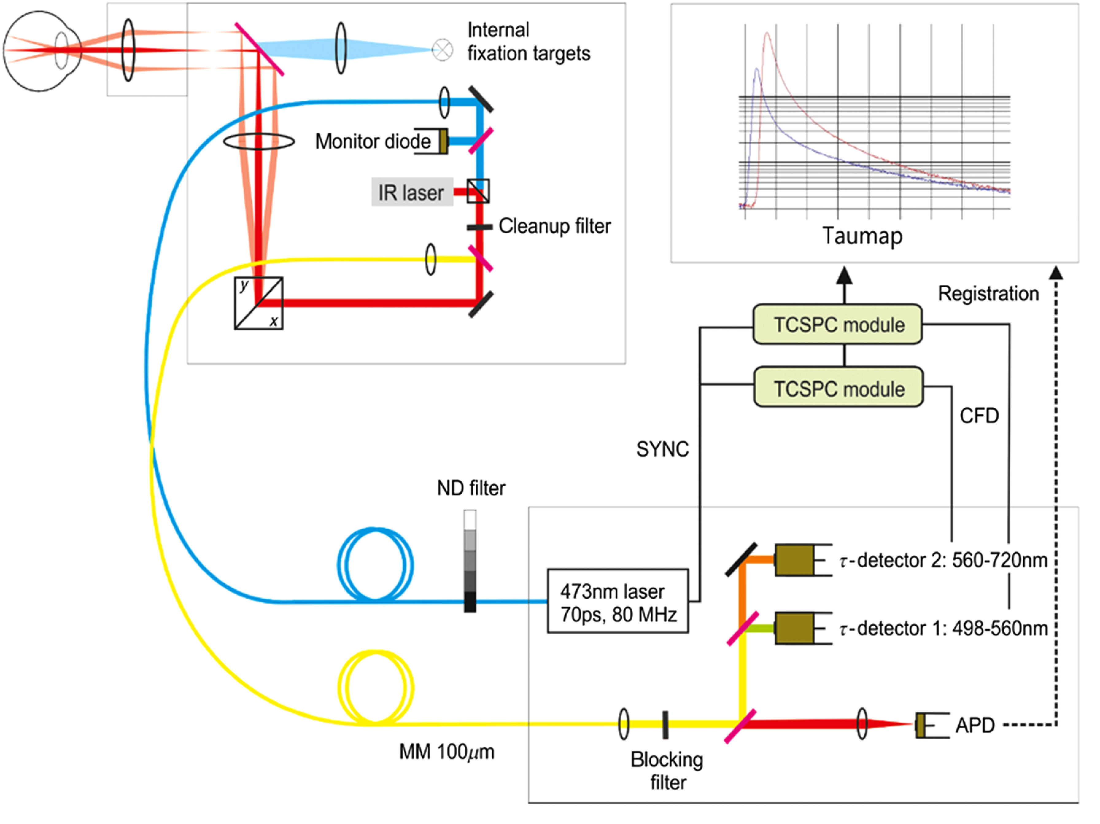
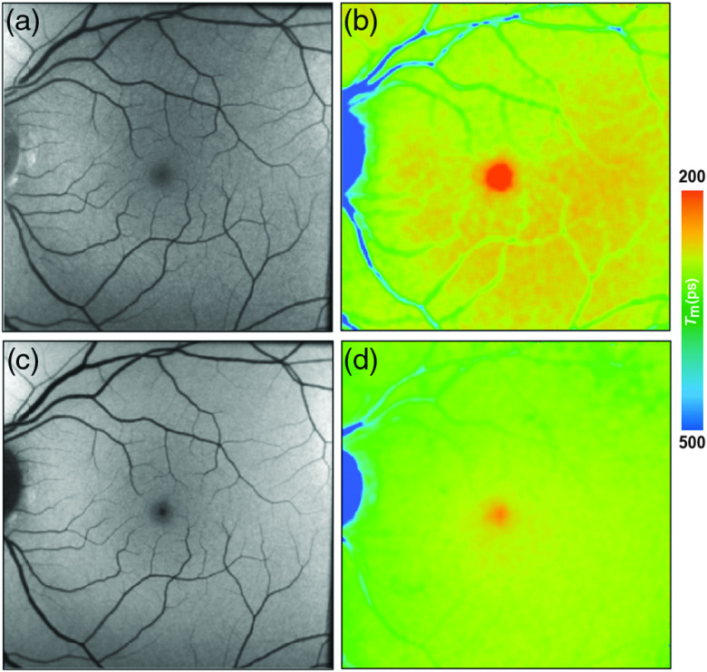
Autofluorescence-based imaging techniques have become very important in the ophthalmological field. Being noninvasive and very sensitive, they are broadly used in clinical routines. Conventional autofluorescence intensity imaging is largely influenced by the strong fluorescence of lipofuscin, a fluorophore that can be found at the level of the retinal pigment epithelium. However, different endogenous retinal fluorophores can be altered in various diseases. Fluorescence lifetime imaging ophthalmoscopy (FLIO) is an imaging modality to investigate the autofluorescence of the human fundus in vivo. It expands the level of information, as an addition to investigating the fluorescence intensity, and autofluorescence lifetimes are captured. The Heidelberg Engineering Spectralis-based fluorescence lifetime imaging ophthalmoscope is used to investigate a 30-deg retinal field centered at the fovea. It detects FAF decays in short [498 to 560 nm, short spectral channel (SSC) and long (560 to 720 nm, long spectral channel (LSC)] spectral channels, the mean fluorescence lifetimes (τm) are calculated using bi- or triexponential approaches. These are meant to be relatively independent of the fluorophore's intensity; therefore, fluorophores with less intense fluorescence can be detected. As an example, FLIO detects the fluorescence of macular pigment, retinal carotenoids that help protect the human fundus from light damages. Furthermore, FLIO is able to detect changes related to various retinal diseases, such as age-related macular degeneration, albinism, Alzheimer's disease, diabetic retinopathy, macular telangiectasia type 2, retinitis pigmentosa, and Stargardt disease. Some of these changes can already be found in healthy eyes and may indicate a risk to developing such diseases. Other changes in already affected eyes seem to indicate disease progression. This review article focuses on providing detailed information on the clinical findings of FLIO. This technique detects not only structural changes at very early stages but also metabolic and disease-related alterations. Therefore, it is a very promising tool that might soon be used for early diagnostics.

Keywords: fluorescence lifetime; fluorescence lifetime imaging ophthalmoscopy; lipofuscin; macular pigment; protein glycation; retinal disease; time-resolved fundus autofluorescence.


Figures

Fig. 1
Fig. 1
The Perrin–Jablonski diagram highlights the photophysical processes involved in fluorescence and phosphorescence.
Fig. 2
Fig. 2
The graph schematically shows the mechanism of a triexponential approximation to obtain the amplitude weighted mean autofluorescence lifetime τm. The fluorescence intensity (y-axis) is depicted over the time t (x-axis). τm is calculated based on the amplitudes and the sum of three exponential functions (components 1 to 3).
Fig. 3
Fig. 3
The basic setup of the scanning fluorescence lifetime imaging ophthalmoscope is depicted. A pulsed 473-nm laser excites retinal fluorescence in a 30-deg field. Fluorescence photons are then detected according to their wavelength in two spectral channels (1: short spectral channel, SSC: 498 to 560 nm; 2: long spectral channel, LSC: 560 to 720 nm). An infrared (IR) laser is used for eye tracking.
Fig. 4
Fig. 4
(a, c) FAF intensity and (b, d) lifetime images from a 25-year-old healthy person. Images of (a, b) the SSC (498 to 560 nm) and (c, d) the LSC (560 to 720 nm) are presented.
Fig. 5
Fig. 5
(a, c) FAF intensity and (b, d) lifetime images from a 44-year-old patient with albinism. Images of (a, b) the SSC (498 to 560 nm) and (c, d) the LSC (560 to 720 nm) are presented.
Fig. 6
Fig. 6
(a, c) FAF intensity and (b, d) lifetime images from a 73-year-old patient with AMD. Images of (a, b) the SSC (498 to 560 nm) and (c, d) the LSC (560 to 720 nm) are presented.
Fig. 7
Fig. 7
(a, c) FAF intensity and (b, d) lifetime images of the SSC (498 to 560 nm) from a 63-year-old patient with DR. Images of (a, b) the SSC (498 to 560 nm) and (c, d) the LSC (560 to 720 nm) are presented.
Fig. 8
Fig. 8
(a, c) FAF intensity and (b, d) lifetime images from a 58-year-old patient MacTel. Images of the (a, b) SSC (498 to 560 nm) and (c, d) the LSC (560 to 720 nm) are presented.
Fig. 9
Fig. 9
(a, c) FAF intensity and (b, d) lifetime images from a patient with a BRAO. Images of (a, b) the SSC (498 to 560 nm) and (c, d) the LSC (560 to 720 nm) are presented.
Fig. 10
Fig. 10
(a, c) FAF intensity and (b, d) lifetime images from a 48-year-old patient with Stargardt disease. Images of (a, b) the SSC (498 to 560 nm) and (c, d) the LSC (560 to 720 nm) are presented. * highlight different flecks.
Fig. 11
Fig. 11
(a, c) FAF intensity and (b, d) lifetime images from a 33-year-old patient with CSCR. Images of (a, b) the SSC (498 to 560 nm) and (c, d) the LSC (560 to 720 nm) are presented.
Fig. 12
Fig. 12
(a, c) FAF intensity and (b, d) lifetime images from a 18-year-old patient with choroideremia. Images of (a, b) the SSC (498 to 560 nm) and (c, d) the LSC (560 to 720 nm) are presented.
Fig. 13
Fig. 13
(a, c) FAF intensity and (b, d) lifetime images from a 45-year-old patient with RP. Images of (a, b) the SSC (498 to 560 nm) and (c, d) the LSC (560 to 720 nm) are presented.

References

    1. Lakowicz J., Principles of Fluorescence Spectroscopy, Plenum Publishers, New York: (1999).
    1. De Los Santos C., et al. , “FRAP, FLIM, and FRET: detection and analysis of cellular dynamics on a molecular scale using fluorescence microscopy,” Mol. Reprod. Dev. 82(7–8), 587–604 (2015).MREDEE10.1002/mrd.22501 - DOI - PMC - PubMed
    1. Chen L. C., et al. , “Fluorescence lifetime imaging microscopy for quantitative biological imaging,” Methods Cell Biol. 114, 457–488 (2013).MCBLAG10.1016/B978-0-12-407761-4.00020-8 - DOI - PubMed
    1. Becker W., et al. , “Fluorescence lifetime imaging by time-correlated single-photon counting,” Microsc. Res. Tech. 63(1), 58–66 (2004).MRTEEO10.1002/jemt.v63:1 - DOI - PubMed
    1. Wang B. G., et al. , “Intraocular multiphoton microscopy with subcellular spatial resolution by infrared femtosecond lasers,” Histochem. Cell Biol. 126(4), 507–515 (2006).10.1007/s00418-006-0187-0 - DOI - PubMed

Publication types