. 2019 Feb;60(2):158-166.
doi: 10.1165/rcmb.2018-0021OC.

The Effects of IFN-λ on Epithelial Barrier Function Contribute to Klebsiella pneumoniae ST258 Pneumonia

Affiliations

The Effects of IFN-λ on Epithelial Barrier Function Contribute to Klebsiella pneumoniae ST258 Pneumonia

Danielle Ahn et al. Am J Respir Cell Mol Biol. 2019 Feb.

Abstract

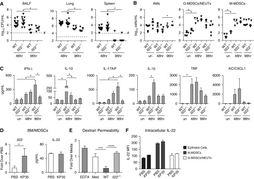
IFN-λ and IL-22, cytokines that share the coreceptor IL-10RB, are both induced over the course of Klebsiella pneumoniae ST258 (KP35) pneumonia. IL-22 is known to protect mucosal barriers, whereas the effects of IFN-λ on the mucosa are not established. We postulated that IFN-λ plays a role in regulating the airway epithelial barrier to facilitate cellular trafficking to the site of infection. In response to IFN-λ, the transmigration of neutrophils across a polarized monolayer of airway epithelial cells was increased, consistent with diminished epithelial integrity. KP35 infection increased epithelial permeability, and pretreatment with IFN-λ amplified this effect and facilitated bacterial transmigration. These effects of IFN-λ were confirmed in vivo, in that mice lacking the receptor for IFN-λ (Ifnlr1-/-) were protected from bacteremia in a murine model of KP35 pneumonia. Conversely, the integrity of the epithelial barrier was protected by IL-22, with subsequent impairment of neutrophil and bacterial transmigration in vitro. Maximal expression of IL-22 in vivo was observed later in the course of infection than IFN-λ production, with high levels of IL-22 produced by recruited immune cells at 48 hours, consistent with a role in epithelial barrier recovery. The divergent and opposing expression of these two related cytokines suggests a regulated interaction in the host response to KP35 infection. A major physiological effect of IFN-λ signaling is a decrease in epithelial barrier integrity, which facilitates immune cell recruitment but also enables K. pneumoniae invasion.

Keywords: IFN-λ; IL-22; airway epithelial barrier; bacterial pneumonia.


Figures

Figure 1.
Figure 1.
Klebsiella pneumoniae alters the expression of epithelial junctional proteins. (A) Ingenuity pathway analysis showing major canonical pathways significantly affected by KP35 infection of wild-type (WT) mice, using proteomic data obtained from pooled BAL fluid (n = 3) after 48 hours of infection. B-H = Benjamini-Hochberg. (B) Heatmaps of the canonical pathways of epithelial remodeling and tight junctional signaling showing relative expression of proteins in KP35-infected mice compared with PBS-treated controls. (C) Immunoblots of ezrin (Ezr), occludin (Ocln), zonula-occludens-1 (ZO-1), and Claudin-2 (Cn-2) from lung homogenates of WT mice infected with KP35 at the indicated time points. Densitometry values normalized to actin (Act) are shown (n = 3); *P < 0.05, **P < 0.01, ***P < 0.005; one-way ANOVA, Dunnett’s post hoc test for multiple comparisons as compared with PBS. For all graphs, each column is the mean value ± SEM. Representative data from at least two independent experiments are shown. (D) Distribution of ZO-1 by immunofluorescence on nonparaffin-fixed lung sections. Lower panels represent magnification of the area defined by the white dashed line boxes in the upper panels. Scale bars: 300 μm.
Figure 2.
Figure 2.
KP35 promotes production of the IL-10RB–related cytokines IFN-λ and IL-22. (A) Heatmaps of the IL-10RB signaling pathway recovered from BAL fluid of KP35-infected mice at 48 hours of infection (same samples as in Figures 1A and 1B; n = 3). (B) Immunoblot with densitometry and qRT-PCR of IL-10RB using lung homogenates of WT mice infected with KP35 at the indicated time points. (C) Gene expression measured via qRT-PCR of lung homogenates for IFN-λ (Ifnl2/3) (n = 6) and IL-22 (Il22) (n = 6), and (D) their respective protein levels measured via ELISA (n = 3) or multiplex array (n = 6) in BAL fluid from mice infected with KP35 at the indicated time points. For immunoblots, densitometry values are normalized to actin (n = 3). Representative data from at least two independent experiments are shown. For all graphs, each column is the mean value ± SEM. *P < 0.05, **P < 0.01, ***P < 0.001, and ****P < 0.0001, by one-way ANOVA; Dunnett’s correction for multiple comparisons between groups indicated by the horizontal bar (B–D).
Figure 3.
Figure 3.
IFN-λ stimulates airway epithelial cells and compromises barrier integrity. (A) Immunoblot and densitometry of confluent human airway epithelial cells (16HBEs) stimulated with their respective cytokine for 24 hours (n = 3). (B and C) qRT-PCR of EZR and OCLN after stimulation of 16HBE cells with IFN-λ (B) or IL-22 (C) for 24 hours (n = 4). (D) qRT-PCR of IFN-λ (IFNL2/3) and IL-22 (IL22) (n = 5) levels in 16HBE cells after 24 hours of KP35 infection. (E and F) Dextran permeability with or without basolateral pretreatment of IFN-λ (E) or IL-22 (F) for 24 hours (n = 8, 2 compiled experiments). (G) The percent change in transepithelial electrical resistance (TEER) before and after treatment with IFN-λ or IL-22 for 24 hours (n = 4). (H) Neutrophil transmigration across polarized 16HBE cells after 4 hours of apical KP35 infection. (I) KP35 cfu (n = 6) recovered from the basolateral side of polarized 16HBE cells after 4 hours of apical infection with or without basolateral pretreatment of IFN-λ or IL-22 for 24 hours (n = 3). EDTA served as a positive control for transmigration assays. For column graphs, the bar is the mean value ± SEM. Representative data from at least two independent experiments are shown, unless otherwise indicated. For all graphs, each column is the mean value ± SEM. *P < 0.05, **P < 0.01, ***P < 0.001, and ****P < 0.0001, by Student’s t test (B–D) or one-way ANOVA, Dunnett’s post hoc test for multiple comparisons compared with media (MED) control (E, F, H, and I) or between the means of every other mean in other columns in the data set. Veh = vehicle.
Figure 4.
Figure 4.
Ifnlr1−/− mice are protected from bacteremia. WT and Ifnlr1−/− mice were infected with KP35 for 48 or 96 hours and compared with PBS-treated controls. (A) Bacterial load was enumerated in BAL fluid (BALF), lung, and spleen. (B) Cell populations in BALF: alveolar macrophages (AMs) (CD45+SiglecF+CDll11blo-mid), granulocytic myeloid-derived suppressor cells/neutrophils (G-MDSCs/NEUTs) (CD45+CD11b+MHCIIloLy6ChiLy6Ghi), and monocytic myeloid-derived suppressor cells (M-MDSCs) (CD45+CD11b+MHCIIloLy6ChiLy6Glo). (C) Cytokines in BALF (n = 8–9). (D) Representative immunoblot of lung homogenate for Ocln and Ezr; densitometry is the result of two experiments compiled (n = 4). For the scatter plots, each point represents an individual mouse and horizontal lines within represent median values. #Represents the limit of detection. For column graphs, the bar is the mean value ± SEM. Compiled data from at least two independent experiments are shown. Horizontal bars above the data sets equal *P < 0.05, **P < 0.01, Kruskal-Wallis test, one-way ANOVA, Dunn’s correction for multiple comparisons. *P < 0.05, **P < 0.01, ***P < 0.001, and ****P < 0.0001. un = untreated.
Figure 5.
Figure 5.
IL-22 increases barrier integrity during KP35 infection. (A) Bacterial load was enumerated in selected compartments. (B) Cell populations in BALF, as defined in Figure 4. (C) Cytokines in BALF from WT and Il22−/− mice (n = 4). (D) Expression of IL-22 via qRT (whole-cell lysate) and ELISA (cell culture supernatant) by BM-MDSCs infected with KP35 compared with PBS controls (n = 4). (E) Dextran permeability of polarized 16HBE cells with or without 24-hour pretreatment with supernatants harvested from WT or Il22−/− BM-MDSCs after 4 hours of KP35 infection. (F) Geometric mean fluorescence intensity of intracellular IL-22 staining of epithelial cells (Epcam+CD11b), M-MDSCs, or neutrophils isolated from lung homogenate of WT mice infected for 48 hours. For scatter plots, each point represents an individual mouse and horizontal lines within represent median values. For column graphs, the bar is the mean value ± SEM. Representative data from at least two independent experiments are shown. *P < 0.05, ***P < 0.005, ****P < 0.001; Kruskal-Wallis test, one-way ANOVA, Dunn’s correction for multiple comparisons; #represents the limit of detection. BM = bone marrow; MFI = mean fluorescence intensity.

References

    1. Gomez-Simmonds A, Greenman M, Sullivan SB, Tanner JP, Sowash MG, Whittier S, et al. Population structure of Klebsiella pneumoniae causing bloodstream infections at a New York City tertiary care hospital: diversification of multidrug-resistant isolates. J Clin Microbiol. 2015;53:2060–2067. - PMC - PubMed
    1. Tumbarello M, Trecarichi EM, De Rosa FG, Giannella M, Giacobbe DR, Bassetti M, et al. ISGRI-SITA (Italian Study Group on Resistant Infections of the Società Italiana Terapia Antinfettiva) Infections caused by KPC-producing Klebsiella pneumoniae: differences in therapy and mortality in a multicentre study. J Antimicrob Chemother. 2015;70:2133–2143. - PubMed
    1. Ahn D, Peñaloza H, Wang Z, Wickersham M, Parker D, Patel P, et al. Acquired resistance to innate immune clearance promotes Klebsiella pneumoniae ST258 pulmonary infection. JCI Insight. 2016;1:e89704. - PMC - PubMed
    1. Moore TA, Perry ML, Getsoian AG, Newstead MW, Standiford TJ. Divergent role of gamma interferon in a murine model of pulmonary versus systemic Klebsiella pneumoniae infection. Infect Immun. 2002;70:6310–6318. - PMC - PubMed
    1. Aujla SJ, Chan YR, Zheng M, Fei M, Askew DJ, Pociask DA, et al. IL-22 mediates mucosal host defense against Gram-negative bacterial pneumonia. Nat Med. 2008;14:275–281. - PMC - PubMed

Publication types

MeSH terms

LinkOut - more resources