

Opposing mechanisms underlying differential changes in brain oxygen and temperature induced by intravenous morphine
- PMID: 30183460
- PMCID: PMC6295537
- DOI: 10.1152/jn.00445.2018
Opposing mechanisms underlying differential changes in brain oxygen and temperature induced by intravenous morphine
Abstract
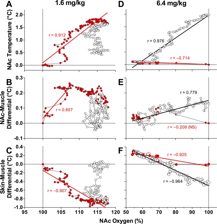
Morphine remains widely used in clinical settings due to its potent analgesic properties. However, one of the gravest risks of all opioids is their ability to induce respiratory depression and subsequent brain hypoxia that can lead to coma and death. Due to these life-threatening effects, our goal was to examine the effects of intravenous morphine at a wide range of doses (0.1-6.4 mg/kg) on changes in brain oxygen levels in freely moving rats. We used oxygen sensors coupled with high-speed amperometry and conducted measurements in the nucleus accumbens (NAc) and subcutaneous (SC) space, the latter serving as a proxy for blood oxygen levels that depend on respiratory activity. We also examined the effects of morphine on NAc, muscle, and skin temperature. Morphine induced dose-dependent decreases in SC oxygen levels, suggesting respiratory depression, but differential effects on NAc oxygen: increases at low and moderate doses (0.1-1.6 mg/kg) and decreases at the highest dose tested (6.4 mg/kg). Morphine also increased brain temperature at low and moderate doses but induced a biphasic, down-up change at high doses. The oxygen increases appear to result from a neurovascular coupling mechanism via local vasodilation and enhanced oxygen entry into brain tissue to compensate for blood oxygen drops caused by modest respiratory depression. At high morphine doses, this adaptive mechanism is unable to compensate for the enhanced respiratory depression, resulting in brain hypoxia. Hence, morphine appears to be safe when used as an analgesic at clinically relevant doses but poses great risks at high doses, likely to be abused by drug users. NEW & NOTEWORTHY With the use of oxygen sensors coupled with amperometry, we show that morphine induces differential effects on brain oxygen levels, slightly increasing them at low doses and strongly decreasing them at high doses. In contrast, morphine dose dependently decreases oxygen levels in the SC space. Therefore, morphine engages opposing mechanisms affecting brain oxygen levels, enhancing them through neurovascular coupling at low, clinically relevant doses and decreasing them due to dramatic respiratory depression at high doses, likely to be abused.
Keywords: metabolism; neurovascular coupling; nucleus accumbens; opioids; oxygen electrochemistry; rats.
Figures


Similar articles
-
Respiratory depression and brain hypoxia induced by opioid drugs: Morphine, oxycodone, heroin, and fentanyl.Neuropharmacology. 2019 Jun;151:219-226. doi: 10.1016/j.neuropharm.2019.02.008. Epub 2019 Feb 5. Neuropharmacology. 2019. PMID: 30735692 Free PMC article. Review.
-
6-Monoacetylmorphine (6-MAM), Not Morphine, Is Responsible for the Rapid Neural Effects Induced by Intravenous Heroin.ACS Chem Neurosci. 2019 Aug 21;10(8):3409-3414. doi: 10.1021/acschemneuro.9b00305. Epub 2019 Jul 8. ACS Chem Neurosci. 2019. PMID: 31268284
-
Intravenous Heroin Induces Rapid Brain Hypoxia and Hyperglycemia that Precede Brain Metabolic Response.eNeuro. 2017 Jun 7;4(3):ENEURO.0151-17.2017. doi: 10.1523/ENEURO.0151-17.2017. eCollection 2017 May-Jun. eNeuro. 2017. PMID: 28593192 Free PMC article.
-
Inflow of oxygen and glucose in brain tissue induced by intravenous norepinephrine: relationships with central metabolic and peripheral vascular responses.J Neurophysiol. 2018 Feb 1;119(2):499-508. doi: 10.1152/jn.00692.2017. Epub 2017 Nov 8. J Neurophysiol. 2018. PMID: 29118201 Free PMC article.
-
Reflections on: "A general role for adaptations in G-Proteins and the cyclic AMP system in mediating the chronic actions of morphine and cocaine on neuronal function".Brain Res. 2016 Aug 15;1645:71-4. doi: 10.1016/j.brainres.2015.12.039. Epub 2015 Dec 29. Brain Res. 2016. PMID: 26740398 Free PMC article. Review.
Cited by
-
Interruption of continuous opioid exposure exacerbates drug-evoked adaptations in the mesolimbic dopamine system.Neuropsychopharmacology. 2020 Oct;45(11):1781-1792. doi: 10.1038/s41386-020-0643-x. Epub 2020 Feb 20. Neuropsychopharmacology. 2020. PMID: 32079024 Free PMC article.
-
No evidence of abnormal metabolic or inflammatory activity in the brains of patients with rheumatoid arthritis: results from a preliminary study using whole-brain magnetic resonance spectroscopic imaging (MRSI).Clin Rheumatol. 2020 Jun;39(6):1765-1774. doi: 10.1007/s10067-019-04923-5. Epub 2020 Jan 30. Clin Rheumatol. 2020. PMID: 32002761 Free PMC article.
-
Machine learning for infection risk prediction in postoperative patients with non-mechanical ventilation and intravenous neurotargeted drugs.Front Neurol. 2022 Aug 1;13:942023. doi: 10.3389/fneur.2022.942023. eCollection 2022. Front Neurol. 2022. PMID: 35979059 Free PMC article.
-
Respiratory depression and brain hypoxia induced by opioid drugs: Morphine, oxycodone, heroin, and fentanyl.Neuropharmacology. 2019 Jun;151:219-226. doi: 10.1016/j.neuropharm.2019.02.008. Epub 2019 Feb 5. Neuropharmacology. 2019. PMID: 30735692 Free PMC article. Review.
References
-
- Benyó Z, Wahl M. Opiate receptor-mediated mechanisms in the regulation of cerebral blood flow. Cerebrovasc Brain Metab Rev 8: 326–357, 1996. - PubMed
-
- Bolger FB, McHugh SB, Bennett R, Li J, Ishiwari K, Francois J, Conway MW, Gilmour G, Bannerman DM, Fillenz M, Tricklebank M, Lowry JP. Characterisation of carbon paste electrodes for real-time amperometric monitoring of brain tissue oxygen. J Neurosci Methods 195: 135–142, 2011. doi:10.1016/j.jneumeth.2010.11.013. - DOI - PubMed
Publication types
MeSH terms
Substances
LinkOut - more resources
Full Text Sources
Other Literature Sources