. 2018 Sep 5;13(9):e0202201.
doi: 10.1371/journal.pone.0202201. eCollection 2018.

Validation of diffusion tensor imaging measures of nigrostriatal neurons in macaques

Affiliations

Validation of diffusion tensor imaging measures of nigrostriatal neurons in macaques

Joshua S Shimony et al. PLoS One. .

Abstract

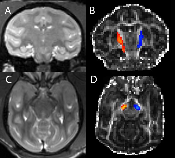
Objective: Interpretation of diffusion MRI in the living brain requires validation against gold standard histological measures. We compared diffusion values of the nigrostriatal tract to PET and histological results in non-human primates (NHPs) with varying degrees of unilateral nigrostriatal injury induced by MPTP, a toxin selective for dopaminergic neurons.

Methods: Sixteen NHPs had MRI and PET scans of three different presynaptic radioligands and blinded video-based motor ratings before and after unilateral carotid artery infusion of variable doses of MPTP. Diffusion measures of connections between midbrain and striatum were calculated. Then animals were euthanized to quantify striatal dopamine concentration, stereologic measures of striatal tyrosine hydroxylase (TH) immunostained fiber density and unbiased stereologic counts of TH stained nigral cells.

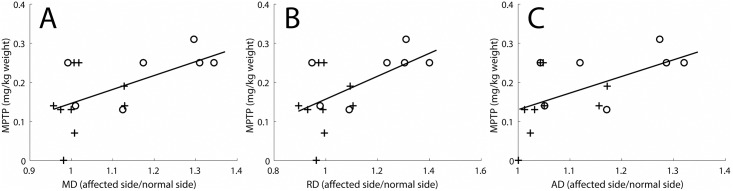
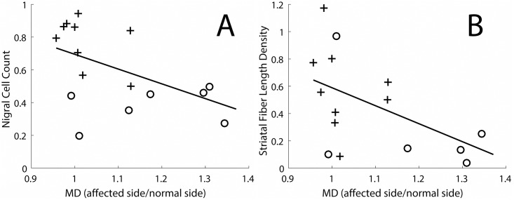
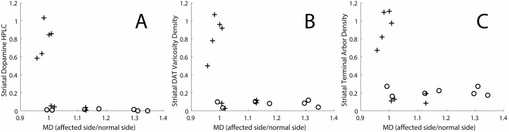
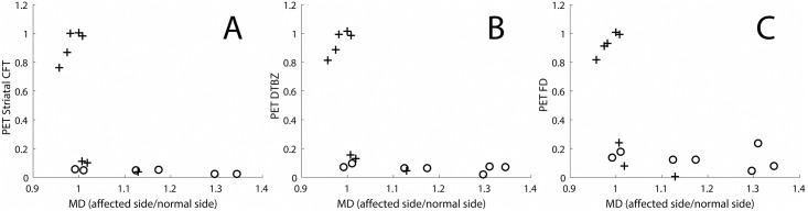
Results: Diffusion measures correlated with MPTP dose, nigral TH-positive cell bodies and striatal TH-positive fiber density but did not correlate with in vitro nigrostriatal terminal field measures or in vivo PET measures of striatal uptake of presynaptic markers. Once nigral TH cell count loss exceeded 50% the stereologic terminal field measures reached a near zero floor effect but the diffusion measures continued to correlate with nigral cell counts.

Conclusion: Diffusion measures in the nigrostriatal tract correlate with nigral dopamine neurons and striatal fiber density, but have the same relationship to terminal field measures as a previous report of striatal PET measures of presynaptic neurons. These diffusion measures have the potential to act as non-invasive index of the severity of nigrostriatal injury. Diffusion imaging of the nigrostriatal tract could potentially have diagnostic value in humans with Parkinson disease or related disorders.


Conflict of interest statement

The authors have declared that no competing interests exist.

Figures

Fig 1
Fig 1. Nigrostriatal track volume.
T2 weighted coronal image through the striatum (A) and FA weighted image (B) with superposed nigrostriatal track volumes (right in orange and left in blue) in a single monkey. Analogous transverse images at a level above the substantia nigra are shown in (C) and (D).
Fig 2
Fig 2. Diffusivity measures correlated with MPTP dose.
Diffusivity ratios (dimensionless, affected side divided by normal side) correlated with MPTP dose (mg/kg body weight). The diffusivity measures include (A) Mean diffusivity (R2 = 0.33, p = 0.02), (B) Radial diffusivity (R2 = 0.31, p = 0.02), and (C) Axial diffusivity (R2 = 0.32, p = 0.02). Crosses indicate monkeys in which there was less than 50% loss of nigral cell bodies, and circles indicate monkeys in which there was a greater than 50% loss of nigral cell bodies.
Fig 3
Fig 3. Nigral cell count and striatal fiber length density as a function of MD in the nigrostriatal tract.
(A) Nigral cell count as a function of the MD in the nigrostriatal tract (R2 = 0.28, p = 0.03). (B) Striatal fiber length density as a function of the MD in the nigrostriatal tract (R2 = 0.29, p = 0.04). Crosses indicate monkeys in which there was less than 50% loss of nigral cell bodies, and circles indicate monkeys in which there was a greater than 50% loss of nigral cell bodies.
Fig 4
Fig 4. Histological measures of terminal fields of nigrostriatal neurons in the striatum as a function of the MD in the nigrostriatal tract.
The measures include (A) HPLC quantification of striatal dopamine measures, (B) Stereological measure of DAT varicosity density, (C) Average terminal arbor density. Crosses indicate monkeys in which there was less than 50% loss of nigral cell bodies, and circles indicate monkeys in which there was a greater than 50% loss of nigral cell bodies.
Fig 5
Fig 5. In-vivo PET measures of pre-synaptic markers as a function of the MD in the nigrostriatal tract.
The measures include (A) Striatal CFT (a DAT marker), (B) DTBZ (a VMAT2 marker), (C) FD (a decarboxylase marker). Crosses indicate monkey’s in which there was less than 50% loss of nigral cell bodies, and circles indicate monkeys in which there was a greater than 50% loss of nigral cell bodies.

References

    1. Johansen-Berg H, Behrens TE. Diffusion MRI London: Elsevier; 2009.
    1. Jones DK, Knosche TR, Turner R. White matter integrity, fiber count, and other fallacies: the do’s and don’ts of diffusion MRI. Neuroimage. 2013;73:239–54. Epub 2012/08/01. 10.1016/j.neuroimage.2012.06.081 . - DOI - PubMed
    1. Karimi M, Tian L, Brown CA, Flores HP, Loftin SK, Videen TO, et al. Validation of nigrostriatal positron emission tomography measures: critical limits. Ann Neurol. 2013;73(3):390–6. Epub 2013/02/21. 10.1002/ana.23798 . - DOI - PMC - PubMed
    1. Perlmutter JS, Norris SA. Neuroimaging biomarkers for Parkinson disease: facts and fantasy. Ann Neurol. 2014;76(6):769–83. Epub 2014/11/05. 10.1002/ana.24291 . - DOI - PMC - PubMed
    1. Tian L, Karimi M, Brown CA, Loftin SK, Perlmutter JS. In vivo measures of nigrostriatal neuronal response to unilateral MPTP treatment. Brain Res. 2014;1571:49–60. Epub 2014/05/23. 10.1016/j.brainres.2014.05.009 . - DOI - PMC - PubMed

Publication types

MeSH terms

Substances