. 2018 Oct 9;18(20):3129-3143.
doi: 10.1039/c8lc00322j.

3D microfluidic ex vivo culture of organotypic tumor spheroids to model immune checkpoint blockade

Affiliations

3D microfluidic ex vivo culture of organotypic tumor spheroids to model immune checkpoint blockade

Amir R Aref et al. Lab Chip. .

Abstract

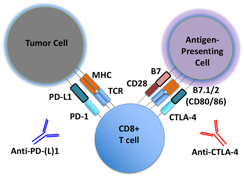
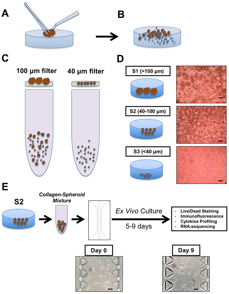
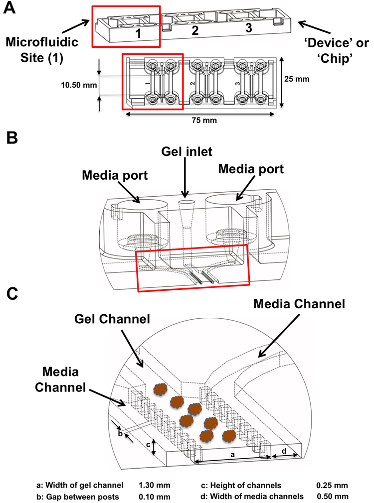
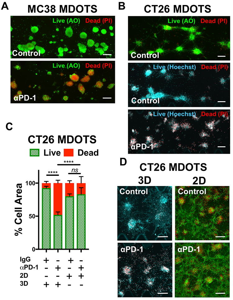
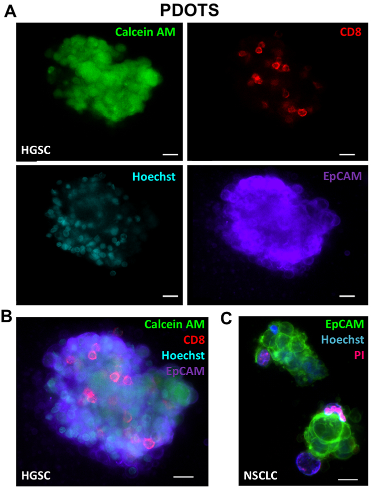
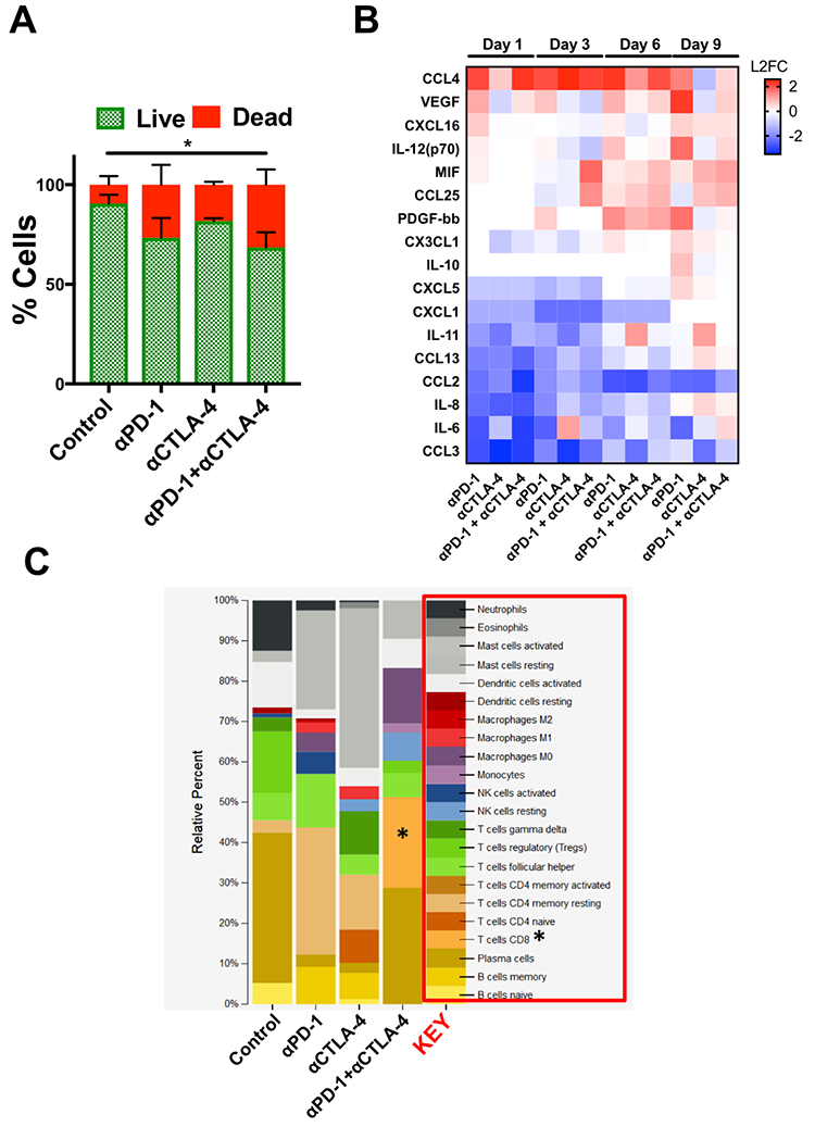
Microfluidic culture has the potential to revolutionize cancer diagnosis and therapy. Indeed, several microdevices are being developed specifically for clinical use to test novel cancer therapeutics. To be effective, these platforms need to replicate the continuous interactions that exist between tumor cells and non-tumor cell elements of the tumor microenvironment through direct cell-cell or cell-matrix contact or by the secretion of signaling factors such as cytokines, chemokines and growth factors. Given the challenges of personalized or precision cancer therapy, especially with the advent of novel immunotherapies, a critical need exists for more sophisticated ex vivo diagnostic systems that recapitulate patient-specific tumor biology with the potential to predict response to immune-based therapies in real-time. Here, we present details of a method to screen for the response of patient tumors to immune checkpoint blockade therapy, first reported in Jenkins et al. Cancer Discovery, 2018, 8, 196-215, with updated evaluation of murine- and patient-derived organotypic tumor spheroids (MDOTS/PDOTS), including evaluation of the requirement for 3D microfluidic culture in MDOTS, demonstration of immune-checkpoint sensitivity of PDOTS, and expanded evaluation of tumor-immune interactions using RNA-sequencing to infer changes in the tumor-immune microenvironment. We also examine some potential improvements to current systems and discuss the challenges in translating such diagnostic assays to the clinic.


Conflict of interest statement

Disclosure of Potential Conflicts

A. Aref, D.A. Barbie, and R.W. Jenkins have ownership interest in pending U.S. Patent Application No. 15/540,346.. R.D. Kamm has ownership interest (including patents) in AIM Biotech, D.A. Barbie is a consultant/advisory board member for N of One.

Figures

Figure 1 –
Figure 1 –. Schematic of PD-1/CTLA-4 Blockade. Schematic detailing basic steps involved in generation of tumor-specific T cells.
Shown is a schematic of a tumor cell, CD8 effector T cell, and an antigen-presenting cell (APC), with associated cell-cell interactions via PD-1/PD-L1 and CTLA-4/B7. Tumor-associated antigens or neo-antigens are presented by major histocompatibility complex (MHC) on APCs or tumor cells to T cells with appropriate T-cell receptor (TCR). CD28 co-activating receptor on T cells binds B7 on APCs. Anti-PD-(L)1 and anti-CTLA-4 antibodies are shown.
Figure 2 –
Figure 2 –. MDOTS/PDOTS Workflow.
(A-B), A tumor specimen is received and subjected to physical and enzymatic dissociation (A), yielding dissociated tumor tissue (B) containing spheroids, single cells, and macroscopic tumor. (C-D), This heterogeneous mixture is then sequentially applied to 100 μm and 40 μm filters (C) to obtain three separate fractions (D), S1 (>100 μm), S2 (40–100 μm), and S3 (<40 μm). E, the S2 fraction is pelleted and resuspended in collagen to be injected into the microfluidic culture device for subsequent ex vivo culture with indicated terminal readouts. Scale bars indicate 100 μm (D, E).
Figure 3 –
Figure 3 –. Microfluidic device.
(A), the 3D cell culture chip (AIM Biotech) is shown with three independent microfluidic chambers per chip. Red rectangle identifies a single microfluidic chamber in the 3D cell culture chip. (B-C), each device contains a center gel region with posts separating the gel region from the anti-parallel side channels. Gel loading port and media ports labeled (B), along with center and side channels (C).
Figure 4 –
Figure 4 –. Live/Dead imaging and analysis of Murine-Derived Organotypic Tumor Spheroids.
(A), acridine orange (AO) and propidium iodide (PI) staining of MC38 MDOTS on Day 6 of ex vivo culture, comparing control (isotype control IgG, 10 μg/mL) with anti-PD-1 (10 μg/mL). B, AO/PI and Hoechst/PI staining of CT26 MDOTS on Day 5 of ex vivo culture, comparing control (isotype control IgG, 10 μg/mL) with anti-PD-1 (10 μg/mL). C-D, Live/Dead analysis (C) and fluorescence images (D) of CT26 MDOTS treated with IgG or anti-PD-1 (10 μg/mL) for 5 days in 3D microfluidic culture (“3D”) compared to 384-well plates (“2D”) (3D - Ho/PI; 2D - AO/PI) (****p<0.0001, ns = not significant; Kruskal-Wallis with multiple comparisons; n≥3). Scale bars indicate 200 μm (A, B, D).
Figure 5 –
Figure 5 –. Fluorescence Imaging of Patient-Derived Organotypic Tumor Spheroids.
(A-B), baseline IF staining of HGSC PDOTS demonstrating viable cells (calcein AM; green), CD8 T cells (red), tumor cells (EpCAM; purple), and all nucleated cells (Hoechst; blue). C, overlay IF image of NSCLC PDOTS demonstrating EpCAM positive tumor cells (green), all nucleated cells (Hoechst; blue), and dead cells (PI; red). Scale bars indicate 20 μm (A, B, C).
Figure 6 –
Figure 6 –. Ex Vivo Profiling of ICB Using PDOTS.
(A), AO/PI staining of SI-NET PDOTS (Day 9) treated with αPD-1, αCTLA-4, and αPD-1 + αCTLA-4 compared to untreated control PDOTS. (B), Heatmap of changes in secreted cytokines from PDOTS (SI-NET); represented as L2FC relative to untreated control at each time point. (C), Inferred changes in PDOTS immune cell populations using CIBERSORT from SI-NET PDOTS RNA-seq.

References

    1. Pardoll DM, Nat Rev Cancer, 2012, 12, 252–264. - PMC - PubMed
    1. Topalian SL, Drake CG and Pardoll DM, Cancer Cell, 2015, 27, 450–461. - PMC - PubMed
    1. Carbognin L, Pilotto S, Milella M, Vaccaro V, Brunelli M, Calio A, Cuppone F, Sperduti I, Giannarelli D, Chilosi M, Bronte V, Scarpa A, Bria E and Tortora G, PLoS One, 2015, 10, e0130142. - PMC - PubMed
    1. Mahoney KM, Rennert PD and Freeman GJ, Nat Rev Drug Discov, 2015, 14, 561–584. - PubMed
    1. Jenkins RW, Barbie DA and Flaherty KT, Br J Cancer, 2018, 118, 9–16. - PMC - PubMed

Publication types