. 2018 Oct 1;10(5):460-474.
doi: 10.1093/jmcb/mjy047.

SUMO-1 modification of FEN1 facilitates its interaction with Rad9-Rad1-Hus1 to counteract DNA replication stress

Affiliations

SUMO-1 modification of FEN1 facilitates its interaction with Rad9-Rad1-Hus1 to counteract DNA replication stress

Xiaoli Xu et al. J Mol Cell Biol. .

Abstract

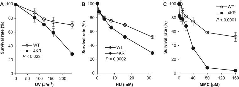
Human flap endonuclease 1 (FEN1) is a structure-specific, multi-functional endonuclease essential for DNA replication and repair. We and others have shown that during DNA replication, FEN1 processes Okazaki fragments via its interaction with the proliferating cell nuclear antigen (PCNA). Alternatively, in response to DNA damage, FEN1 interacts with the PCNA-like Rad9-Rad1-Hus1 complex instead of PCNA to engage in DNA repair activities, such as homology-directed repair of stalled DNA replication forks. However, it is unclear how FEN1 is able to switch between these interactions and its roles in DNA replication and DNA repair. Here, we report that FEN1 undergoes SUMOylation by SUMO-1 in response to DNA replication fork-stalling agents, such as UV irradiation, hydroxyurea, and mitomycin C. This DNA damage-induced SUMO-1 modification promotes the interaction of FEN1 with the Rad9-Rad1-Hus1 complex. Furthermore, we found that FEN1 mutations that prevent its SUMO-1 modification also impair its ability to interact with HUS1 and to rescue stalled replication forks. These impairments lead to the accumulation of DNA damage and heightened sensitivity to fork-stalling agents. Altogether, our findings suggest an important role of the SUMO-1 modification of FEN1 in regulating its roles in DNA replication and repair.


Figures

Figure 1
Figure 1
Identification and validation of FEN1 SUMO-1 modification. (A) Western blot analysis was used to detect SUMOylated and unmodified FEN1 in UV-treated (120 J/m2, 3-h recovery) HeLa cells treated with siRNA targeting the coding region of Ubc9 gene (si-Ubc9) or a scrambled siRNA control (–). Cells were harvested 3 h post-UV irradiation. FEN1 proteins in whole cell extracts were detected using an antibody against human FEN1. Relative protein levels of Ubc9 and β-actin (loading control) were detected using anti-Ubc9 and anti-β-actin antibodies. (B) FEN1 is SUMO-1 modified in HeLa cells in response to UV treatment. UV treatment was conducted as described for Panel 1A, and cells were harvested at 3 h post-UV irradiation. FEN1 was isolated from whole cell extracts via IP, and FEN1 and SUMO-1-FEN1 were detected by western blot analysis using antibodies against FEN1 and SUMO-1, respectively. β-actin was used as a control to ensure equal input for each IP reaction. To accurately reflect the amount of FEN1 in different samples, we could not overexpose the immunoblot. Therefore, the SUMO-1-FEN1 was not shown here. (C) Exogenously expressed 3×FLAG-tagged FEN1 (FLAG-FEN1) is SUMO-1-modified after UV treatment. HeLa cells stably expressing FLAG-FEN1 were treated with siRNA targeting the 5′UTR of Ubc9 gene si-Ubc9 or negative scrambled siRNA control. To restore Ubc9 expression, HeLa cells were co-transfected with si-Ubc9 and a vector encoding human Ubc9. FLAG-FEN1 was immunoprecipitated using anti-FLAG M2 beads. SUMO-1-FLAG-FEN1 and FLAG-FEN1 were detected by western blot using anti-SUMO-1 or FEN1 antibodies. Ubc9 levels were verified using an anti-Ubc9 antibody. β-actin was used as a loading control. (D) SUMO-1 modification of recombinant FEN1 in vitro is Ubc9-mediated. Purified recombinant FEN1 as incubated with Ubc9 and SUMO-1 for 60 min at 37°C. Unmodified FEN1 and SUMO-1-FEN1 were visualized using Coomassie Brilliant Blue staining and western blot analysis using antibodies against FEN1 and SUMO-1. (E) HeLa cells stably expressing 3×FLAG-tagged FEN1 were exposed to UV irradiation and allowed to recover for 0, 2, 4, or 6 h. Cells not exposed to UV irradiation were used as controls (CON). Cells were harvested and total 3×FLAG-FEN1 was isolated via IP. 3×FLAG-FEN1 and SUMO-1-3×FLAG-FEN1 were detected by western blot using anti-FEN1 or anti-SUMO-1 antibodies. The top panel shows the representative western blot images, and the bottom panel shows the quantification of SUMO-1-FEN1 relative to levels in UV-unexposed control cells at 0 h. The intensity of SUMO-1-3×FLAG-FEN1 bands in the SUMO-1 blot was normalized to the corresponding 3×FLAG-FEN1 band in the FLAG blot. Values shown are mean ± SD of three independent assays. P-values were calculated using Student’s t-test for each time point. ns, not significant, *P < 0.05. (F) HeLa cells stably expressing 3×FLAG-tagged FEN1 were exposed to UV irradiation (120 J/m2, 3-h recovery) or treated with HU (1 mM, 3 h) or MMC (18 μM, 3 h). FEN1 was purified from treated cells and untreated controls using anti-FLAG M2 magnetic beads, and 3×FLAG-FEN1 and SUMO-1-3×FLAG-FEN1 were detected by western blot analysis using anti-FEN1 and anti-SUMO-1 antibodies.
Figure 2
Figure 2
K366, K367, K369, and K375 residues are the primary SUMO-1 modification sites of FEN1. (A) Purified FLAG-tagged WT or mutant (K366R, K367R, K369R, K375R, or 4KR) FEN1 proteins were incubated with SUMO-1 and SUMO-1 modification reaction components. FEN1 and SUMO-1-FEN1 were detected by western blot analysis using anti-FLAG and anti-SUMO-1 antibodies. The quantified intensities of SUMO-1 modification of the mutant FEN1 proteins, normalized to corresponding 3×FLAG FEN1 levels and relative to that of WT FEN1, are shown. (B) WT or 4KR FEN1 were incubated with SUMO-1 modification reaction components, with or without SUMO-1. FEN1 and SUMO-1-FEN1 levels were detected in a single blot using an anti-FEN1 antibody. (C) HeLa cells stably expressing 3×FLAG-tagged WT or 4KR mutant FEN1 were exposed to UV (120 J/m2, 3-h recovery) and 3×FLAG-tagged WT or 4KR FEN1 was purified with anti-FLAG M2 magnetic beads. 3×FLAG-tagged FEN1 and SUMO-1-3×FLAG-FEN1 were detected by western blot analysis. (D) HeLa cells stably expressing 3×FLAG-tagged WT or 4KR mutant FEN1 were exposed to UV irradiation (120 J/m2, 3-h recovery) or treated with HU (1 mM, 3 h), CPT (5 μM, 3 h), or MMC (18 μM, 3 h). 3×FLAG-tagged WT and 4KR mutant FEN1 were purified with anti-FLAG M2 beads, and 3×FLAG-FEN1 and SUMO-1-3×FLAG-FEN1 were detected by western blot analysis. The intensities of SUMO-1-FEN1, normalized to corresponding 3×FLAG FEN1 levels and relative to that of untreated WT FEN1, are shown. (E) SUMO-1 modification of WT and 4KR FEN1 was visualized in HeLa cells using the Duolink®in situ PLA with anti-SUMO-1 and anti-FLAG antibodies (PLA-SUMO-1/3×FLAG). Nuclei were stained with DAPI. Scale bars: 10 μm.
Figure 3
Figure 3
Phosphorylation of FEN1 stimulates its SUMO-1 modification. (A) Various FEN1 modifications were analyzed in HeLa cells expressing 3×FLAG-FEN1 with or without UV irradiation (120 J/m2, 3-h recovery). 3×FLAG-FEN1 was purified using M2 beads. Methylated (Me-FEN1), phosphorylated (Phos-FEN1), and SUMO-1-modified FEN1 were detected by western blot analysis using anti-FLAG, anti-methylarginine, anti-phosphoserine, and anti-SUMO-1 antibodies. β-actin served as a control to ensure equal input for each IP reaction. (B) HeLa cells expressing 3×FLAG-FEN1 were treated with the CDK inhibitor olomoucine (40 μM, 24 h) or a vehicle control (DMSO) and exposed to UV irradiation. FLAG-FEN1, SUMO-FEN1, and Phos-FEN1 were detected, as in Panel A. GAPDH served as a control to ensure equal input for each IP reaction. Left panel: representative western blot images. Right panel: Quantification of phosphorylated and SUMO-1 modified FEN1. The intensities of Phos-FEN1 or SUMO-1-FEN1 bands, normalized to the corresponding 3×FLAG-FEN1 band and relative to that of UV-unexposed cells, are shown. Values shown are mean ± SD of three independent assays. P-values were calculated using Student’s t-test. *P < 0.05, **P < 0.01. (C) HeLa cells were transfected with myc-tagged WT, S187A phosphorylation-defective, or S187D phosphorylation-mimic FEN1. Myc-tagged WT, S187A, or S187D FEN1 was isolated using myc beads. SUMO-1-myc-FEN1 (WT or mutant) was detected using western blot analysis with an anti-SUMO-1 antibody. FEN1 levels were determined using anti-myc and anti-FEN1 antibodies. GAPDH served as a control to ensure equal input for each IP reaction. (D) SUMO-1 modification of purified WT, S187A, and S187D FEN1 was conducted in vitro. ATP was or was not added to initiate the reaction. Anti-FEN1 and anti-SUMO-1 antibodies were used to detect FEN1 and SUMO-1-FEN1.
Figure 4
Figure 4
SUMOylation of FEN1 facilitates its interaction with RAD1 and HUS1. (A) In vitro binding assays were conducted for FEN1 or SUMO-1-FEN1 with PCNA or HUS1. Purified recombinant 6×His-tagged FEN1 was incubated with Ubc9, SUMO-1, and other components of the SUMOylation kit, with or without ATP, at 37°C for 60 min. Unmodified FEN1 or SUMO-1-FEN1 was incubated with Ni-NTA beads. After extensive washing, the beads were incubated with a mixture of MBP-tagged PCNA and GST-tagged HUS1 proteins. Incubation of PCNA and HUS1 with beads only (no FEN1) and incubation of FEN1-Ni-NTA beads with non-conjugated MBP and GST proteins without ATP served as negative controls for non-specific binding of the proteins or tags to the beads. FEN1-bound MBP-PCNA and GST-HUS1 were analyzed by western blot analysis using anti-PCNA, anti-HUS1, anti-MBP, and anti-GST antibodies. The intensities of PCNA and HUS1 in the pull-down were quantified and normalized to their input levels. The relative levels of PCNA and HUS1 binding to FEN1, with versus without SUMO-1 modification (ATP), are shown. (B) WT HeLa cells were treated with 120 J/m2 UV irradiation and allowed to recover for 3 h. FEN1 complexes were immunoprecipitated using an anti-FEN1 antibody. FEN1 and SUMO-1-FEN1, as well as PCNA, HUS1, RAD1 that were co-IPed with FEN1, were detected by western blot analysis. A bead-only (no anti-FEN1) control was used as a negative control for IP. The relative levels of PCNA, HUS1, and RAD1 binding to FEN1, normalized to the loading control IgG and relative to that of untreated WT cells, are shown. (C) HeLa cells stably expressing 3×FLAG-tagged WT or 4KR mutant FEN1 were treated with 120 J/m2 UV irradiation and allowed to recover for 3 h. 3×FLAG-FEN1 complexes were immunoprecipitated using anti-FLAG M2 beads. 3×FLAG-FEN1 and SUMO-1-FEN1, as well as PCNA, HUS1, and RAD1 that were co-immunoprecipitated with FEN1, were detected by western blot analysis. (D) The interactions of WT or 4KR mutant FEN1 with PCNA, HUS1, and RAD1 with and without UV treatment were analyzed using the Duolink®in situ PLA with antibody mixtures containing anti-FLAG/anti-PCNA, anti-FLAG/anti-HUS1, and anti-FLAG/anti-RAD-1. Nuclei were stained with DAPI. Upper panels show representative PLA assay images (scale bars: 10 μm). and the bottom panels show PLA intensities, relative to that of control WT cells. Values shown are mean ± SD of three independent assays. P-values were calculated using Student’s t-test. ns, not significant, *P < 0.05, **P < 0.01.
Figure 5
Figure 5
Nuclease activities of unmodified- and SUMO-1-modified FEN1. (A and B) FEN activity on double flap substrates (A) and GEN activity on gapped duplex substrates (B) were assessed in vitro. Purified FEN1 was incubated for 60 min with the SUMO-1 modification kit reaction mixture at 37°C in the absence or presence of ATP. The resultant unmodified (no ATP control) FEN1 or SUMO-1-FEN1 was then incubated with FAM-labeled DNA substrates in reaction buffer at 37°C for 5, 10, 15, 20, or 25 min (duration indicated by the wedge above the gel). The reactions were resolved in a 15% denaturing PAGE gel and visualized with a Typhoon FLA 9500 imager.
Figure 6
Figure 6
SUMOylation of FEN1 affects its DNA damage repair activity in vivo and in vitro. (A) WT or 4KR mutant HeLa cells were pulse-labeled with the thymidine analog IdU (25 μM) for 30 min, treated with HU (4 mM) or DMSO for 4 h, and then pulse-labeled with the thymidine analog CIdU (125 μM) for 40 min. The cells were lysed and the DNA fibers were spread, and stained using anti-IdU and anti-CIdU antibodies. Representative images show stalled DNA forks that failed to restart (green), newly firing forks (red), and restarted forks (green/red). (B) Percentages of each type of replication fork (restarted, non-restarted, and newly firing) in WT and 4KR cells with or without HU treatment. Values are mean ± SD of three independent assays. (C) DNA replication and proliferation of WT and 4KR mutant HeLa cells were analyzed by EdU incorporation after cells were synchronized to S phase. Representative microimages show EdU-positive cells in WT and 4KR cells with or without UV irradiation (120 J/m2 UV, 3-h recovery). Nuclei were stained with DAPI (Hoechst blue), EdU incorporation (green) marks cells undergoing proliferation. Scale bars: 60 μm for the images with low magnification and 10 μm for the enlarged images. (D) Percentages of EdU-positive WT and 4KR cells with or without UV irradiation. Values are mean ± SD of three assays. P-values were calculated using Student’s t-test. *P < 0.05.
Figure 7
Figure 7
FEN1 SUMOylation-defective cells accumulate DNA damage. (A and B) Representative immunofluorescence images of DNA damage markers γH2AX and 53BP1 in the nuclei of WT and 4KR mutant HeLa cells. Cells were stained using anti-γH2AX and anti-53BP1 antibodies. Scale bars: 20 μm. (C) Representative immunofluorescence images of γH2AX and 53BP1 in the nuclei of WT of 4KR cells after UV irradiation (120 J/m2 UV, 3-h recovery) or treated with HU (1 mM, 3 h) or MMC (18 μM, 3 h). Scale bars: 30 μm. (D) Quantification of γH2AX- and 53BP1-positive nuclei under each DNA damaging condition. Values are mean ± SD of three independent assays. P-values were calculated using Student’s t-test. *P < 0.05, **P < 0.01. (E) Western blot analysis using anti-γH2AX and anti-53BP1 antibodies in WT and 4KR cells with or without exposure to UV irradiation. β-actin was used as a loading control.
Figure 8
Figure 8
Defective SUMO-1 modification of FEN1 impairs cell cycle progression and sensitizes cells to replication fork-stalling agents. Survival rates of WT and 4KR mutant HeLa cells, determined using MTS cell proliferation assays after treatment with varying levels of UV irradiation (A) or HU (B) or MMC (C) treatment, are shown. Survival rates were calculated as the absorbance 490 (A490) of a sample divided by the A490 of the corresponding untreated control, multiplied by 100%. Values are mean ± SD of three independent assays. P-values, indicated in each panel, were calculated using ANOVA test.

Similar articles

Cited by

References

    1. Anderson L., Henderson C., and Adachi Y. (2001). Phosphorylation and rapid relocalization of 53BP1 to nuclear foci upon DNA damage. Mol. Cell. Biol. 21, 1719–1729. - PMC - PubMed
    1. Aravind L., Walker D.R., and Koonin E.V. (1999). Conserved domains in DNA repair proteins and evolution of repair systems. Nucleic Acids Res. 27, 1223–1242. - PMC - PubMed
    1. Ayyagari R., Gomes X.V., Gordenin D.A., et al. . (2003). Okazaki fragment maturation in yeast. I. Distribution of functions between FEN1 and DNA2. J. Biol. Chem. 278, 1618–1625. - PubMed
    1. Bae S.H., Bae K.H., Kim J.A., et al. . (2001). RPA governs endonuclease switching during processing of Okazaki fragments in eukaryotes. Nature 412, 456–461. - PubMed
    1. Bai H., Madabushi A., Guan X., et al. . (2010). Interaction between human mismatch repair recognition proteins and checkpoint sensor Rad9-Rad1-Hus1. DNA Repair (Amst) 9, 478–487. - PMC - PubMed

Publication types