

In vitro evaluation of anti-fibrotic effects of select cytokines for vocal fold scar treatment
- PMID: 30184328
- PMCID: PMC7011756
- DOI: 10.1002/jbm.b.34198
In vitro evaluation of anti-fibrotic effects of select cytokines for vocal fold scar treatment
Abstract
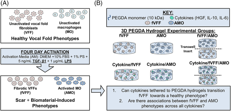
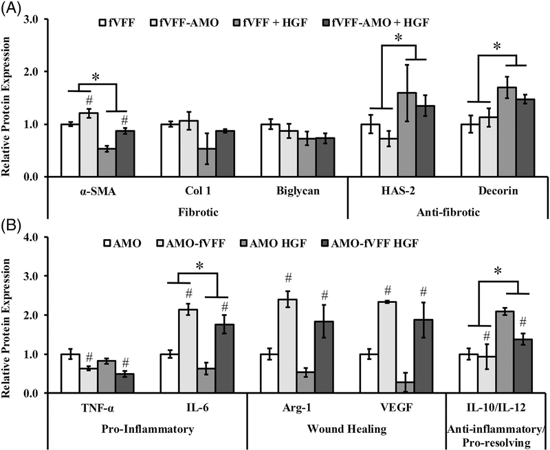
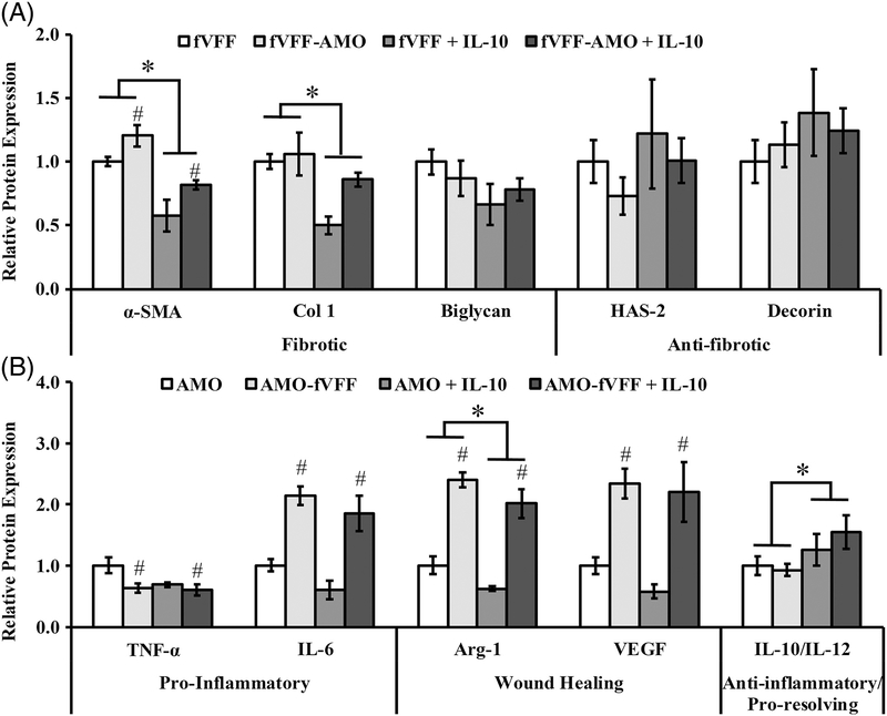
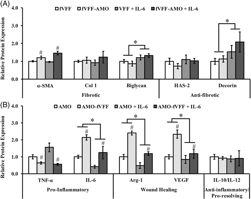
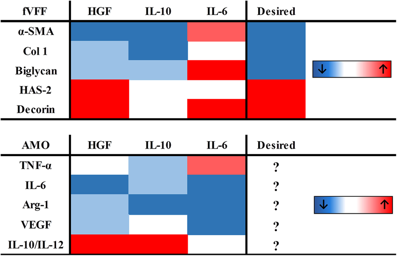
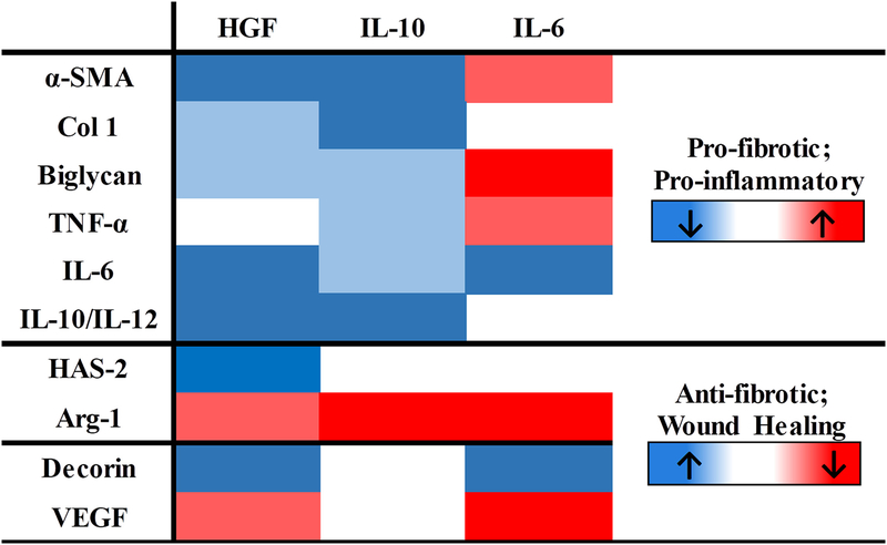
Scarring of the vocal fold lamina propria (LP) can cause considerable voice disorders due to reduced pliability in scar tissue, attributed in part to abnormal extracellular matrix (ECM) deposition produced by the fibrotic vocal fold fibroblast (fVFF). Cytokines with anti-fibrotic potential have been investigated to limit abnormal LP ECM, but are limited by the need for repeat injections. Moreover, the potentially significant role played by activated macrophages (AMOs) is usually not considered even though the interaction between AMO and fibrotic fibroblasts is known to regulate scar formation across different tissues. AMO are also regulated by cytokines that are used for LP scar removal, but little is known about AMO behaviors in response to these cytokines within the context of LP scar. In the present study, we evaluated anti-fibrotic effects of hepatocyte growth factor (HGF), interleukin-10 (IL-10) and interleukin-6 (IL-6) in a 3D, in vitro fVFF-AMO co-culture system using poly(ethylene glycol) diacrylate (PEGDA) hydrogels. Data from all cytokines was synthesized into a heat-map that enabled assessment of specific associations between AMO and fVFF phenotypes. Cumulatively, our results indicated that both HGF and IL-10 are potentially anti-fibrotic (reduction in fibrotic markers and enhancement in normal, anti-fibrotic VFF markers), while IL-6 displays more complex, marker specific effects. Possible associations between AMO and fVFF phenotypes were found and may highlight a potential desirable macrophage phenotype. These data support the therapeutic potential of HGF and IL-10 for LP scar treatment, and shed light on future strategies aimed at targeting specific AMO phenotypes. © 2018 Wiley Periodicals, Inc. J Biomed Mater Res Part B: Appl Biomater 107B: 1056-1067, 2019.
Keywords: 3D cell culture; cytokine treatment; fibroblasts; macrophages; vocal fold scar.
© 2018 Wiley Periodicals, Inc.
Figures
Similar articles
-
Pathophysiology of Fibrosis in the Vocal Fold: Current Research, Future Treatment Strategies, and Obstacles to Restoring Vocal Fold Pliability.Int J Mol Sci. 2019 May 24;20(10):2551. doi: 10.3390/ijms20102551. Int J Mol Sci. 2019. PMID: 31137626 Free PMC article. Review.
-
In vitro evaluation of a basic fibroblast growth factor-containing hydrogel toward vocal fold lamina propria scar treatment.J Biomed Mater Res B Appl Biomater. 2018 Apr;106(3):1258-1267. doi: 10.1002/jbm.b.33936. Epub 2017 Jun 5. J Biomed Mater Res B Appl Biomater. 2018. PMID: 28580765 Free PMC article.
-
The efficacy of a novel collagen-gelatin scaffold with basic fibroblast growth factor for the treatment of vocal fold scar.J Tissue Eng Regen Med. 2017 May;11(5):1598-1609. doi: 10.1002/term.2060. Epub 2015 Jun 29. J Tissue Eng Regen Med. 2017. PMID: 26119035
-
Modeling fibrosis using fibroblasts isolated from scarred rat vocal folds.Lab Invest. 2016 Jul;96(7):807-16. doi: 10.1038/labinvest.2016.43. Epub 2016 Apr 25. Lab Invest. 2016. PMID: 27111284 Free PMC article.
-
The Role of Cytokines in Modulating Vocal Fold Fibrosis: A Contemporary Review.Laryngoscope. 2021 Jan;131(1):139-145. doi: 10.1002/lary.28507. Epub 2020 Apr 15. Laryngoscope. 2021. PMID: 32293731 Review.
Cited by
-
The Challenges of OSCC Diagnosis: Salivary Cytokines as Potential Biomarkers.J Clin Med. 2020 Sep 4;9(9):2866. doi: 10.3390/jcm9092866. J Clin Med. 2020. PMID: 32899735 Free PMC article. Review.
-
Changes of pro-inflammatory and anti-inflammatory macrophages after peripheral nerve injury.RSC Adv. 2020 Oct 22;10(64):38767-38773. doi: 10.1039/d0ra06607a. eCollection 2020 Oct 21. RSC Adv. 2020. PMID: 35518415 Free PMC article.
-
Pathophysiology of Fibrosis in the Vocal Fold: Current Research, Future Treatment Strategies, and Obstacles to Restoring Vocal Fold Pliability.Int J Mol Sci. 2019 May 24;20(10):2551. doi: 10.3390/ijms20102551. Int J Mol Sci. 2019. PMID: 31137626 Free PMC article. Review.
-
The Modulation of Fibrosis in Vocal Fold Repair: A Study on c-Met Agonistic Antibodies and Hepatocyte Growth in Animal Studies.Medicina (Kaunas). 2024 Dec 10;60(12):2033. doi: 10.3390/medicina60122033. Medicina (Kaunas). 2024. PMID: 39768913 Free PMC article.
-
In Vitro Evaluation of Biomaterials for Vocal Fold Injection: A Systematic Review.Polymers (Basel). 2021 Aug 6;13(16):2619. doi: 10.3390/polym13162619. Polymers (Basel). 2021. PMID: 34451158 Free PMC article. Review.
References
-
- Benninger MS, Alessi D, Archer S, Bastian R, Ford C, Koufman J, Sataloff RT, Spiegel JR, Woo P. Vocal fold scarring: current concepts and management. Otolaryngol Head Neck Surg 1996;115(5): 474–482. - PubMed
-
- Hansen JK, Thibeault SL. Current understanding and review of the literature: vocal fold scarring. J Voice 2006;20(1):110–120. - PubMed
-
- Graupp M, Bachna-Rotter S, Gerstenberger C, Friedrich G, Fröhlich-Sorger E, Kiesler K, Gugatschka M. The unsolved chapter of vocal fold scars and how tissue engineering could help us solve the problem. Eur Arch Oto-Rhino-Laryngol 2016;273(9):2279–2284. - PubMed
-
- Rousseau B, Hirano S, Chan RW, Welham NV, Thibeault SL, Ford CN, Bless DM. Characterization of chronic vocal fold scarring in a rabbit model. J Voice 2004;18(1):116–124. - PubMed
Publication types
MeSH terms
Substances
Grants and funding
LinkOut - more resources
Full Text Sources
Other Literature Sources
Medical
