. 2018 Oct 2;57(39):5738-5747.
doi: 10.1021/acs.biochem.8b00817. Epub 2018 Sep 19.

Molecular Insights into Human Hereditary Apolipoprotein A-I Amyloidosis Caused by the Glu34Lys Mutation

Affiliations

Molecular Insights into Human Hereditary Apolipoprotein A-I Amyloidosis Caused by the Glu34Lys Mutation

Isabel Morgado et al. Biochemistry. .

Abstract

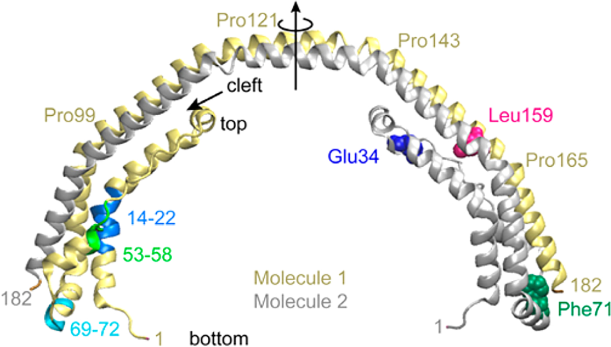
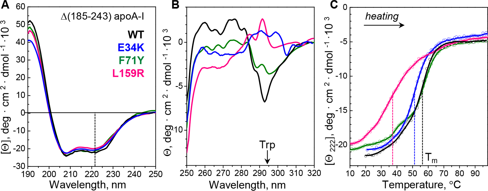
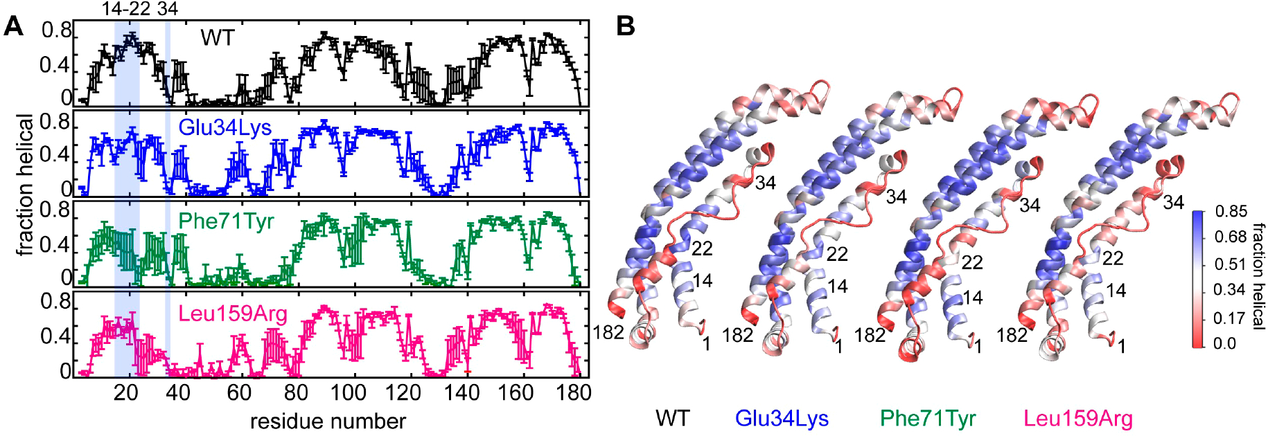
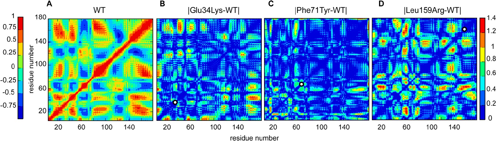
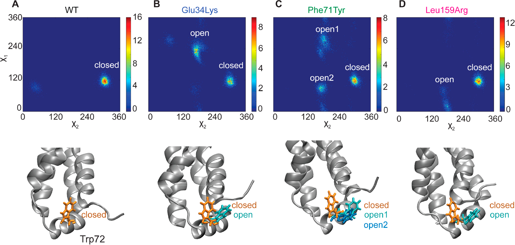
Hereditary apolipoprotein A-I (apoA-I) amyloidosis is a life-threatening incurable genetic disorder whose molecular underpinnings are unclear. In this disease, variant apoA-I, the major structural and functional protein of high-density lipoprotein, is released in a free form, undergoes an α-helix to intermolecular cross-β-sheet conversion along with a proteolytic cleavage, and is deposited as amyloid fibrils in various organs, which can cause organ damage and death. Glu34Lys is the only known charge inversion mutation in apoA-I that causes human amyloidosis. To elucidate the structural underpinnings of the amyloidogenic behavior of Glu34Lys apoA-I, we generated its recombinant globular N-terminal domain (residues 1-184) and compared the conformation and dynamics of its lipid-free form with those of two other naturally occurring apoA-I variants, Phe71Tyr (amyloidogenic) and Leu159Arg (non-amyloidogenic). All variants showed reduced structural stability and altered aromatic residue packing. The greatest decrease in stability was observed in the non-amyloidogenic variant, suggesting that amyloid formation is driven by local structural perturbations at sensitive sites. Molecular dynamics simulations revealed local helical unfolding and suggested that transient opening of the Trp72 side chain induced mutation-dependent structural perturbations in a sensitive region, including the major amyloid hot spot residues Leu14-Leu22. We posit that a shift from the "closed" to the "open" orientation of the Trp72 side chain modulates structural protection of amyloid hot spots, suggesting a previously unknown early step in the protein misfolding pathway.


Conflict of interest statement

The authors declare no competing financial interest.

Figures

Figure 1.
Figure 1.
Atomic structure of the globular domain of lipid-free human apoA-I. The 2.2 Å resolution X-ray crystal structure of free Δ(184–243) WT (Protein Data Bank entry 3R2P) shows a crystallographic dimer comprised of two helix bundles. Dimer molecules 1 (yellow) and 2 (gray) are related by a 2-fold symmetry axis (vertical arrow) that passes through the middle of the central linker (residues 121–142). Domain swapping around this flexible linker is thought to mediate monomer-to-dimer interconversion in apoA-I., In the monomer (shown in Figure 3), all segments in the four-helix bundle are from the same molecule. The top and bottom of the bundle are indicated. A short arrow points to the hydrophobic cleft between two pairs of helices, which is proposed to open upon lipid binding. Mutated side chains explored in this study are shown in molecule 2 in a spherical representation: Glu34 (blue), Phe71 (green), and Leu159 (pink). Amyloid hot spots are shown in molecule 1 in residue segments 14–22 (blue), 53–58 (light green), and 69–72 (cyan).,
Figure 2.
Figure 2.
Conformation and stability of recombinant C-terminally truncated proteins. Globular domains (residues 1–184) of WT (black), Glu34Lys (E34K, blue), Phe71Tyr (F71Y, green), and Leu159Arg (L159R, pink) were obtained and analyzed as described in Materials and Methods. (A) Far-UV CD spectra of the four proteins. Each spectrum represents three to five independent measurements. The helix content assessed from the CD signal at 222 nm (dashed line) ranged from 56 ± 5% in Leu159Arg to 62 ± 5% in WT. (B) Near-UV CD spectra of the four proteins show large differences, particularly at wavelengths dominated by Trp (a peak centered at ~295 nm, shown by an arrow). Each spectrum represents an average of three independent measurements with five-point adjacent averaging. (C) Melting data recorded by CD at 222 nm, Θ222(T), monitor α-helical unfolding during heating. Circles show raw data points. Dashed lines indicate melting temperatures, Tm, corresponding to the first-derivative maxima, dΘ222(T)/dT.
Figure 3.
Figure 3.
Variations in the local helical conformation of WT, Glu34Lys, Phe71Tyr, and Leu159Arg proteins determined from high-temperature MD simulations. (A) Helical fraction vs residue number in apoA-I variants. WT and variant apoA-I are color-coded. For each protein, the dominant structure from the room-temperature simulations was heated to 500 K over 50 ps, followed by simulation for 20 ns at 500 K; the last 10 ns of these simulations was used to determine the helical fraction in each position. Standard errors of three replicates are shown by bars. The area shaded in blue depicts the major amyloidogenic hot spot residues, Leu14–Leu22, and the mutation site Glu34, as indicated by residue numbers at the top. (B) The starting structure for each protein is colored on the basis of the average helicity, from high to low helical fractions (blue to red, respectively), as indicated by the colored bar.
Figure 4.
Figure 4.
Correlation maps illustrate concerted molecular motions in WT, Glu34Lys, Phe71Tyr, and Leu159Arg proteins. The maps were calculated over three independent high-temperature MD simulations. (A) Correlated molecular motions in WT depict protein groups that move either in concert (correlation coefficients from 0 to 1, green to red, respectively) or out of phase (0 to −1, teal to blue, respectively), as indicated in the left color bar. (B–D) Absolute difference between correlation maps of the three variants and WT apoA-I illustrating mutational effects on molecular motion. Groups whose relative motions remained invariant upon mutation are colored blue, and those whose correlated motions changed upon mutation are shown in warm colors (indicated in the right bar). The position of each apoA-I variant is marked with a white dot on the diagonal of each plot.
Figure 5.
Figure 5.
Conformational distribution of the Trp72 side chain in WT and variant proteins. Probability distributions for dihedral angles χ1 and χ2 of Trp72 (top) and structural models depicting the corresponding conformations of the Trp72 side chain (bottom) in (A) WT, (B) Glu34Lys, (C) Phe71Tyr, and (D) Leu159Arg proteins. One representative result of three independent room-temperature simulations is shown. The probability is proportional to the number of steps in the simulation trajectory. Least and most probable conformations are colored blue and red, respectively, according to the color bars on the right. The color bars in different panels are different to illustrate the full range of probabilities for each protein. WT shows the sharpest peak corresponding to the “closed” Trp72 conformation. Glu34Lys, Phe71Tyr, and Leu159Arg proteins show additional peaks corresponding to “open” Trp72 conformations, which are depicted in the bottom panels. Stick models show “closed” (orange) and “open” (cyan and blue) orientations of the Trp72 side chain.

References

    1. Nichols WC, Dwulet FE, Liepnieks J, and Benson MD (1988) Variant apolipoprotein AI as a major constituent of a human hereditary amyloid. Biochem. Biophys. Res. Commun. 156, 762–768. - PubMed
    1. Teoh CL, Griffin MD, and Howlett GJ (2011) Apolipoproteins and amyloid fibril formation in atherosclerosis. Protein Cell 2, 116–127. - PMC - PubMed
    1. Das M, and Gursky O (2015) Amyloid-forming properties of human apolipoproteins: Sequence analyses and structural insights. Adv. Exp. Med. Biol. 855, 175–211. - PMC - PubMed
    1. Rosenson RS, Brewer HB Jr., Davidson WS, Fayad ZA, Fuster V, Goldstein J, Hellerstein M, Jiang XC, Phillips MC, Rader DJ, Remaley AT, Rothblat GH, Tall AR, and Yvan-Charvet L (2012) Cholesterol efflux and atheroprotection: advancing the concept of reverse cholesterol transport. Circulation 125, 1905–1919. - PMC - PubMed
    1. Sorci-Thomas MG, Zabalawi M, Bharadwaj MS, Wilhelm AJ, Owen JS, Asztalos BF, Bhat S, and Thomas MJ (2012) Dysfunctional HDL containing L159R ApoA-I leads to exacerbation of atherosclerosis in hyperlipidemic mice. Biochim. Biophys. Acta, Mol. Cell Biol. Lipids 1821, 502–512. - PMC - PubMed

Publication types

LinkOut - more resources