Review
. 2019 Jul:91:153-168.
doi: 10.1016/j.semcdb.2018.08.015. Epub 2018 Sep 20.

Recapitulating kidney development: Progress and challenges

Affiliations
Review

Recapitulating kidney development: Progress and challenges

Melissa H Little et al. Semin Cell Dev Biol. 2019 Jul.

Abstract

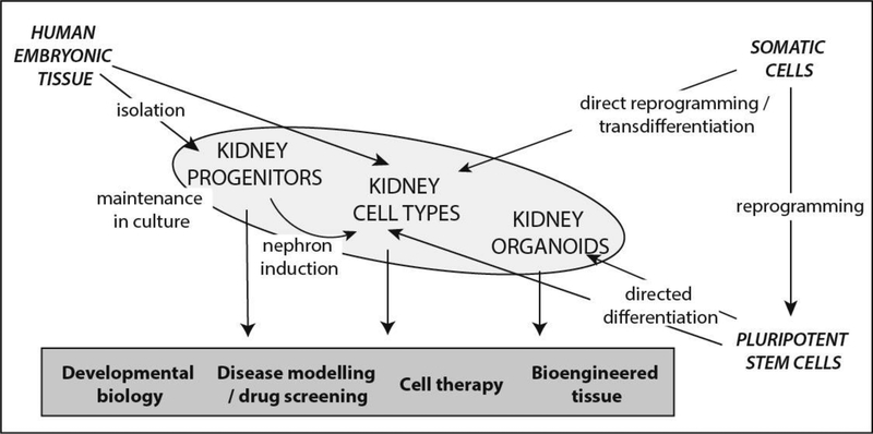
Decades of research into the molecular and cellular regulation of kidney morphogenesis in rodent models, particularly the mouse, has provided both an atlas of the mammalian kidney and a roadmap for recreating kidney cell types with potential applications for the treatment of kidney disease. With advances in both our capacity to maintain nephron progenitors in culture, reprogram to kidney cell types and direct the differentiation of human pluripotent stem cells to kidney endpoints, renal regeneration via cellular therapy or tissue engineering may be possible. Human kidney models also have potential for disease modelling and drug screening. Such applications will rely upon the accuracy of the model at the cellular level and the capacity for stem-cell derived kidney tissue to recapitulate both normal and diseased kidney tissue. In this review, we will discuss the available cell sources, how well they model the human kidney and how far we are from application either as models or for tissue engineering.

Keywords: Directed differentiation; Human nephron progenitor; Kidney development; Kidney organoid; Pluripotent stem cell; Transdifferentiation.


Figures

Figure 1:
Figure 1:
Summary of the sources of human cells and tissues available for studying kidney development, disease modelling and renal regeneration.
Figure 2.
Figure 2.. Published protocols for the directed differentiation of human pluripotent stem cells to kidney endpoints.
Stages of differentiation: AIM, anterior intermediate mesoderm; IM, intermediate mesoderm, MM, metanephric mesenchyme; ND, nephric duct; PIM, posterior intermediate mesoderm; PS, primitive streak; PPS, posterior primitive streak; Tx, transplantation; UB, ureteric bud. Compounds added: A, Activin A; A10, activing A 10uM, B4, BMP4; B7, BMP7; C or CHIR, CHIR990201 (GSK3β antagonist / canonical Wnt activator); C1, CHIR 1uM; C3, CHIR 3uM, C5, CHIR 5uM, C8, CHIR 8uM; F1, FGF1; F2, FGF2; F9, FGF9; G, GDNF; LDN, LDN193189 (BMP Type 1 receptor (ALK2/3) inhibitor); Ng, Noggin (BMP inhibitor), RA, retinoic acid; SB, SB431542 (TGFβ Type 1 receptor (ALK5) inhibitor; Y, Y27632 (Rho-kinase inhibitor).
Figure 3.
Figure 3.. Glomerular vascularisation and tubular maturation post in vivo organoid transplantation under the renal capsule.
A. Visualisation of blood flow (FITC-Dextran) through the glomerulus of an hPSC-derived human kidney organoids generated using a CRISPR-Cas9 gene edited iPSC line in which a blue fluorescent protein has been inserted into the MAFB gene locus in order to mark podocytes. B. TEM image of a proximal tubular segment within an hPSC-derived kidney organoid 2 weeks after transplantation under the renal capsule. Ec, endothelial cell; ptc, peritubular capillary; te, tubular epithelium C,D. Low and higher magnification TEM images of a glomerular basement membrane present between podocytes and endothelial cells of the organoid glomerulus. aj, adherens junction; cap, capillary; ec, endothelial cell; fp, foot process; gbm, glomerular basement membrane. Images courtesy of van den Berg et al [80].
Figure 4.
Figure 4.. Application of hPSC-derived kidney tissues for disease modelling.
A. Outline for the generation of patient-derived and isogenic iPSC lines from a Maizner-Saldino syndrome patient carrying compound heterozygote mutations in the IFT140 gene [107]. The protocol for simultaneous generation of patient and gene corrected lines is described in Howden et al [79]. B. Approach to the characterisation of disease related changes using cells isolated from hPSC-derived kidney organoids [107]. C. Immunofluorescence imaging of renal tubular epithelial cells within iPSC-derived kidney organoids showing the presence of clubbed cilia in patient organoids and wildtype cilia in organoids from gene corrected lines. D. Bright field images of intact glomeruli isolated by sieving dissociated hPSC-derived kidney organoids at low and high power. Figure courtesy of Dr. Lorna Hale.
Figure 5.
Figure 5.. Scale, structure and function represent key challenges to ‘rebuilding a kidney’ using hPSC-derived models.
Images of organoids courtesy of Ms. Pei Xuan Er and Dr. Jessica Vanslambrouck, Murdoch Children’s Research Institute. Image of female human kidney (35 weeks gestation) courtesy of Prof. Mary Jane Black and Dr, Megan Sutherland, Monash University.

References

    1. USRDS. Chapter 6: Healthcare Expenditures for Persons with CKD. USRDS Annual Data Report 2017. [cited 2018.
    1. AIHW, Australia’s Health 2014. 2014, Australian Institute of Health and Welfare: Canberra: p. 178.
    1. Caskey F, Steenkamp R, and Thomas K, UK Renal Registry 19th Annual Report: Introduction. Nephron, 2017. 137(suppl 1)(Suppl. 1): p. 1–10. - PubMed
    1. Cowan PJ, Cooper DK, and d’Apice AJ, Kidney xenotransplantation. Kidney Int, 2014. 85(2): p. 265–75. - PMC - PubMed
    1. Hammerman MR, Xenotransplantation of embryonic pig kidney or pancreas to replace the function of mature organs. J Transplant, 2011. 2011: p. 501749. - PMC - PubMed

Publication types