Review
. 2018 Sep 4;28(3):337-352.
doi: 10.1016/j.cmet.2018.08.014.

The Role of Advanced Glycation End Products in Aging and Metabolic Diseases: Bridging Association and Causality

Affiliations
Review

The Role of Advanced Glycation End Products in Aging and Metabolic Diseases: Bridging Association and Causality

Jyotiska Chaudhuri et al. Cell Metab. .

Abstract

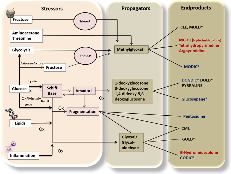
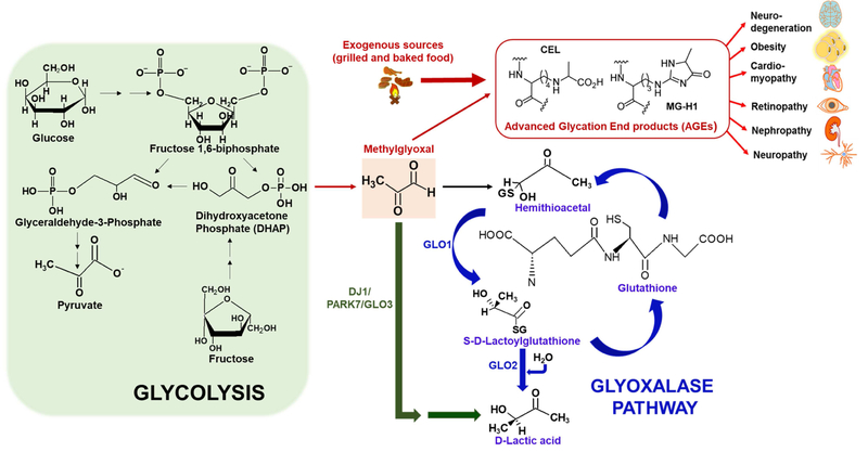
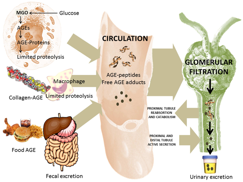
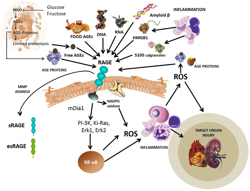
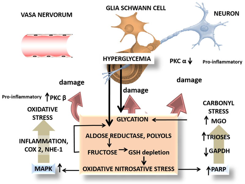
Accumulation of advanced glycation end products (AGEs) on nucleotides, lipids, and peptides/proteins are an inevitable component of the aging process in all eukaryotic organisms, including humans. To date, a substantial body of evidence shows that AGEs and their functionally compromised adducts are linked to and perhaps responsible for changes seen during aging and for the development of many age-related morbidities. However, much remains to be learned about the biology of AGE formation, causal nature of these associations, and whether new interventions might be developed that will prevent or reduce the negative impact of AGEs-related damage. To facilitate achieving these latter ends, we show how invertebrate models, notably Drosophila melanogaster and Caenorhabditis elegans, can be used to explore AGE-related pathways in depth and to identify and assess drugs that will mitigate against the detrimental effects of AGE-adduct development.


Conflict of interest statement

Declaration of Interests

The authors declare no competing interests.

Figures

Figure 1.
Figure 1.. The Maillard reaction or glycation.
Main pathways and main known advanced glycation end products (AGEs) relevant to human pathology. In the left column, we represent the main initiators or stressors, propagators in the center (very potent alpha dicarbonyls, of which methylglyoxal is the most important and reactive) and end products in the right column. Among the endproducts: black represents lysine modifications; red indicates arginine adducts and blue denotes lysine-arginine modifications. CEL: carboxy ethyl lysine; CML: carboxy methyl lysine; MG-H1: methylglyoxal hydroimidazolone; MODIC: methylglyoxal dimer imidazolone crosslink; MOLD: Methylglyoxal lysine dimer; GODIC:glyoxal-derived imidazolium cross-link; DOGDIC, 3-deoxyglucosone-derived imidazolium cross-link; GOLD: glyoxal lysine dimer; DOLD: deoxyglucosone lysine dimer. * indicates crosslinked products. Modified from: (Monnier et al., 2005).
Figure 2.
Figure 2.. Form ation of AGEs and the glyoxalase system.
The figure illustrates the formation and detoxification of MGO, a glycolytic byproduct that is formed either from glucose or fructose. Endogenously derived glycolytic byproducts (e.g., Methylglyoxal or MGO) or their AGE derivatives (e.g., Nε-carboxyethyl-lysine or CEL and Methylglyoxal-derived hydroimidazolone or MG-H1) lead to a variety of diseases affecting different organs (such as brain, heart, eyes, kidney, lungs) that complicate with age. In addition to endogenous sources such as glucose or fructose derived dihydroxyacetone phosphate (DHAP) (formed during glycolysis), exogenous dietary sources such as dry heat-cooked food predominantly produces AGEs (large red arrow). Detoxification of MGO occurs via glutathione (GSH)-dependent glyoxalase pathway mediated by two mitochondrial enzymes glyoxalase 1 (GLO1) and glyoxalase 2 (GLO2) that eventually converts MGO to lactic acid. Alternatively, MGO can also be converted to lactic acid in a single step mediated by a GSH-independent cytosolic enzyme glyoxalase 3 (GLO3), a mammalianorthologue of the protein DJ1/PARK7.
Figure 3.
Figure 3.. AGE m etabolism.
The diagram represents schematically the production of endogenous AGEs (intra and extracellular) and the intake from certain foods (left), their presence in the circulation (center) and their elimination/catabolism mainly by the kidneys (right). AGEs can be produced intracellularly by multiple pathways as shown in Figure 1. Of special relevance are MGO-generated AGEs. Other AGEs are generated on collagens and other proteins in the ECM as we AGE and more rapidly during diabetes. Partial proteolysis by macrophages, or in cells by ubiquitin-mediated proteasome pathways produce partially digested AGE-peptides as well as free adducts. Some AGEs are thought to also enter the bloodstream from foods. These adducts are very reactive and can damage proteins as they transit in the bloodstream. Renal function is critical for their elimination, as end-stage renal failure patients have very high concentrations of these molecules, which can be lowered by dialysis. These AGEs are filtered and reabsorbed in part in the proximal tube to be detoxified to some extent and then secreted distally for urinary excretion.
Figure 4.
Figure 4.. The receptor for advanced glycation endp ro ducts (RAGE) is akey pathway for inflammatory complication sinaging and chronic disease.
As shown in the top part of the figure, RAGE is a pattern recognition receptor that participates in primary immunity and has a variety of ligands. Important for our topic are the AGE, AGE-proteins, AGE-peptides and adducts from primary AGE catabolism and dietary AGEs. RAGE activation leads to a signaling cascade that produces NF-kB coordinated inflammatory responses that can lead to target tissue injury. Soluble forms of the receptor (esRAGE and sRAGE) are released to the circulation. Some authors suggest they serve to modulate the response acting as decoy ligands. They are useful as biomarkers of the whole-body AGE-RAGE axis.
Figure 5:
Figure 5:. Glycation and diabetic neuropathy: an example of pathways and targets where AGEs, MGO, and carbonyl stress play an important role.
Evidence shows that a triple hit may be operating to produce the final damage: vasa vasorum, the Schwann cell and the neuron axon all three are targets for the damage. Hyperglycemia produces AGEs via the mechanisms shown here and in detail in Figure 1. The interplay with oxidative stress (depletion of glutathione) activates MAPK and its inflammatory cascades which via PKC beta produces vascular damage. Oxidative stress on DNA induces the repair enzyme PARP which has the drawback of inactivating GAPDH, therefore blocking glycolysis at the triose level with the upstream consequence of further MGO accumulation, compounding the problem (Brownlee’s hypothesis, see text). Final damage occurs to the neuronal axon, neuronal mitochondria, and the glial Schwann cells.

References

    1. Agalou S, Ahmed N, Babaei-Jadidi R, Dawnay A, and Thornalley PJ (2005). Profound mishandling of protein glycation degradation products in uremia and dialysis. J. Am. Soc. Nephrol 16,1471–1485. - PubMed
    1. Ahmed N, Ahmed U, Thornalley PJ, Hager K, Fleischer G, and Münch G (2005). Protein glycation, oxidation and nitration adduct residues and free adducts of cerebrospinal fluid in Alzheimer’s disease and link to cognitive impairment. J. Neurochem 92, 255–263. - PubMed
    1. Andersson D. a, Gentry C, Light E, Vastani N, Vallortigara J, Bierhaus A, Fleming T, and Bevan S (2013). Methylglyoxal evokes pain by stimulating TRPA1. PLoS One 8, e77986. - PMC - PubMed
    1. Ariga H, Takahashi-Niki K, Kato I, Maita H, Niki T, and Iguchi-Ariga SMM (2013). Neuroprotective function of dj-1 in Parkinson’s disease. Oxid. Med. Cell. Longev - PMC - PubMed
    1. Baba SP, Barski OA, Ahmed Y, O’Toole TE, Conklin DJ, Bhatnagar A, and Srivastava S (2009). Reductive metabolism of AGE precursors: A metabolic route for preventing AGE accumulation in cardiovascular tissue. Diabetes 58, 2486–2497. - PMC - PubMed

Publication types

Substances