. 2018 Sep 5;37(1):219.
doi: 10.1186/s13046-018-0895-z.

The putative tumour suppressor miR-1-3p modulates prostate cancer cell aggressiveness by repressing E2F5 and PFTK1

Affiliations

The putative tumour suppressor miR-1-3p modulates prostate cancer cell aggressiveness by repressing E2F5 and PFTK1

Sen-Mao Li et al. J Exp Clin Cancer Res. .

Abstract

Background: Previous studies report that miR-1-3p, a member of the microRNA-1 family (miR-1), and functions as a tumor suppressor in several different cancers. However, little is known regarding the biological role and intrinsic regulatory mechanisms of miR-1-3p in prostate cancer (PCa).

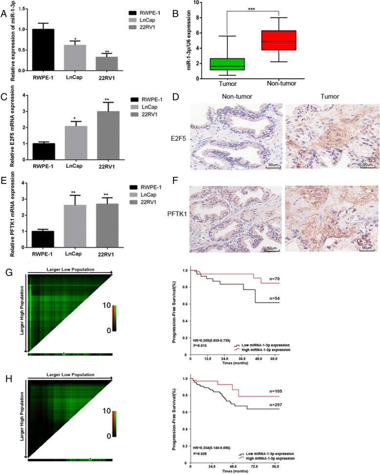
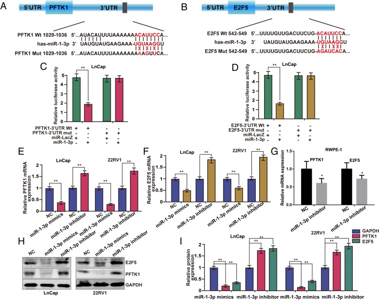
Methods: In this study, the expression levels of miR-1-3p were first examined in PCa cell lines and tumor tissues by RT-qPCR and bioinformatics. The in vitro and in vivo functional effect of miR-1-3p was examined further. A luciferase reporter assay was conducted to confirm target associations.

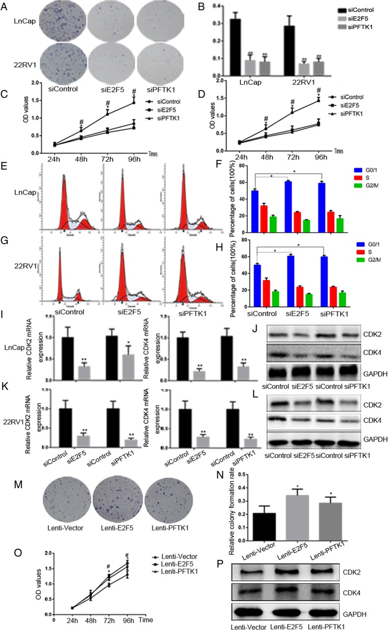
Results: We found that miR-1-3p was significantly downregulated in advanced PCa tissues and cell lines. Low miR-1-3p levels were strongly associated with aggressive clinicopathological features and poor prognosis in PCa patients. Ectopic expression of miR-1-3p in 22RV1 and LncaP cells was sufficient to prevent tumor cell growth and cell cycle progression in vitro and in vivo. Further mechanistic studies revealed that miR-1-3p could directly target the mRNA 3'- untranslated region (3'- UTR) of two central cell cycle genes, E2F5 and PFTK1, and could suppress their mRNA and protein expression. In addition, knockdown of E2F5 and PFTK1 mimicked the tumor-suppressive effects of miR-1-3p overexpression on PCa progression. Conversely, concomitant knockdown of miR-1-3p and E2F5 and PFTK1 substantially reversed the inhibitory effects of either E2F5 or PFTK1 silencing alone.

Conclusion: These data highlight an important role for miR-1-3p in the regulation of proliferation and cell cycle in the molecular etiology of PCa and indicate the potential for miR-1-3p in applications furthering PCa prognostics and therapeutics.

Keywords: Proliferation; Prostate cancer; Target gene; microRNA.


Conflict of interest statement

Ethics approval and consent to participate

All protocols were approved by the Ethics Committee of Tongji Hospital, Tongji Medical College, Huazhong University of Science and Technology, and informed consent was obtained from all patients before surgery.

Consent for publication

Not applicable.

Competing interests

The authors declare that they have no competing interests.

Publisher’s Note

Springer Nature remains neutral with regard to jurisdictional claims in published maps and institutional affiliations.

Figures

Fig. 1
Fig. 1
Mature miR-1-3p expression levels in prostate cancer cell lines and tissues, and the prognostic value of miR-1-3p levels in patients with PCa. a Expression levels of miR-1-3p, (c) E2F5 and (e) PFTK-1 were examined by RT-qPCR in RWPE-1 cells and two PCa cell lines. GAPDH and U6 served as corresponding loading controls. Error bars represent the mean ± S.D. of three independent experiments (*P < 0.05, **P < 0.01 and ***P < 0.001 compared to RWPE-1 group). b Expression levels of mature miR-1-3p in 25 paired PCa and adjacent non-tumour tissues. Alteration of expression is shown as box plot presentations, with the y axis indicating miR-1-3p expression. The mean level of miR-1-3p expression in PCa tissues were significantly lower than that in non tumor tissues. (***P < 0.001, independent t test). d E2F5 and (f) PFTK-1 protein expression in primary prostate tissues was detected by Immunohistochemically staining assay. Scale bar: 50 μm. g 124 PCa patients from the Tongji Hospital, Tongji Medical College, Huazhong University of Science and Technology, and (h) 402 PCa patients from the TCGA database. Left panel: X-tile plots automatically selected the cut-off point of miR-1-3p. Right panel: Kaplan–Meier analysis of survival and the COX proportional hazards model for the hazard ratio
Fig. 2
Fig. 2
MiR-1-3p attenuates prostate cancer cell lines proliferation through inducing cell cycle arrest at G0/G1 phase. LnCap and 22RV1 cells were transfected with 100 nM indicated RNAs molecules respectively for 72 h. a and b MTS assays revealed cell growth curves of both PCa cells in every 24 h. c Representative micrographs and (d) relative quantification of crystal violet-stained cell colonies analyzed by clonogenic formation. e and f flow cytometric determination of proportion of LnCap and 22RV1 cells in distinct cell cycle phases. g and h Expression of CDK2 and CDK4 mRNA in LnCap and 22RV1 cells were assessed by RT-qPCR following transfection with 100 nm miR-1-3p mimics, miR-1-3p inhibitor, or their negative controls (NC). GAPDH served as a loading control. i Western blot analysis of the relative expression of CDK2 and CDK4 in response to indicated RNAs molecules. GAPDH served as a loading control. The results were plotted as the mean ± S.D. of three independent experiments. (*P < 0.05, **P < 0.01, ***P < 0.001, # P < 0.05 and ## P < 0.01)
Fig. 3
Fig. 3
E2F5 and PFTK-1 were identified as direct target genes of miR-1-3p and down-regulated by miR-1-3p in the prostate cancer cell. a and b Schematic diagram of the predicted target binding sites of miR-1-3p in the 3′-UTR of E2F5 and PFTK-1. The seed recognition site is denoted. All nucleotides of the 3′-UTR region of E2F5 and PFTK-1 that binds with miR-1-3p are highly conserved across species as predicated by TargetScan (http://www.targetscan.org/vert_72/). c and d The luciferase activity of the wild type E2F5/ PFTK-1 3’-UTR (Wt) and mutant E2F5/ PFTK-1 3’-UTR (Mut) co-transfected with miR-1-3p mimics or a miRNA negative control (miR-LacZ) was measured in LnCap cells. Relative luciferase activity was plotted as the mean ± S.D. of three independent experiments. e-g Expression of PFTK-1 and E2F5 mRNA in LnCap, 22RV1 and RWPE-1 cells were assessed by RT-qPCR following transfection with miR-1-3p mimics, miR-1-3p inhibitor, or their negative controls (NC). GAPDH served as a loading control. h and i Western blot analysis of the protein levels and relative expression of E2F5 and PFTK-1 in response to 100 nM of indicated RNAs molecules. GAPDH served as a loading control in LnCap and 22RV1 cells. (*P < 0.05, **P < 0.01)
Fig. 4
Fig. 4
E2F5 and PFTK1 are involved to promote PCa cell proliferation and cell cycle progression in vitro. LnCap and 22RV1 cells were transfected with siRNAs of E2F5 and PFTK-1 respectively for 72 h; siControl served as negative control. a Representative photographs of colony formation assay and (b) quantification of the cell colonies formation were used to determine the proliferative ability of PCa Cells. c and d MTS assays revealed cell growth curves of indicated cells in every 24 h. e and f Flow cytometric determination of proportion of indicated cells in distinct cell cycle phases. g-j RT-qPCR and Western blot analysis the gene and protein expression of CDK2 and CDK4. LnCap cells were infected by lenti-E2F5 and PFTK1 to overexpress E2F5/PFTK1. Lenti-vector served as negative control. k and l The cell colony formation was used to determine the proliferative ability. m MTS assays revealed cell growth curves in every 24 h. n Western blot analysis the expression of CDK2 and CDK4 indicated cells. The results were plotted as the mean ± SEM of three independent experiments, with at least three replicates in each independent experiment. (*P < 0.05, **P < 0.01; #P < 0.05, ##P < 0.01)
Fig. 5
Fig. 5
E2F5 and PFTK1 are a functional targets of miR-1-3p suppression of PCa cells proliferation and cell cycle progression. LnCap cells were co-transfected with miR-1-3p inhibitor and siRNA of E2F5 and PFTK-1 respectively for 72 h, inhibitor-NC and siControl served as respective negative control. a The effect of concomitant knockdown of miR-1-3p and E2F5 or PFTK1 on LnCap cells growth rates as measured by MTS assays. b and c Western blot analysis of E2F5 and PFTK1 protein levels in indicated cells. GAPDH was used as the loading control. d The effect of concomitant knockdown of miR-1-3p and E2F5 or PFTK1 on LnCap cells proliferative ability as determined by colony formation assays. e-f The proportion of indicated cells in distinct cell cycle phases as identified by FACS. Results were plotted as the mean ± SEM of three independent experiments, with at least three replicates in each independent experiment. (*P < 0.05, **P < 0.01)
Fig. 6
Fig. 6
MiR-1-3p inhibits PCa tumor xenograft growth in vivo. a Representative photograph of tumor formation and (b) tumors excised 40 days after inoculation of stably transfected cells into nude mice. c Tumor volume was measured using a Vernier caliper on the indicated days. d Tumor weight in the miR-NC and miR-1-3p treated groups. e The relative expression of miR-1-3p in xenograft tumor tissue were identifed by RT-qPCR. f Immunohistochemical analysis of Ki-67,E2F5 and PFTK1 in xenografts tumors of miR-NC and miR-1-3p treated groups. Scale bar: 100 μm. (*P < 0.05, **P < 0.01)

Similar articles

Cited by

References

    1. He L, Hannon GJ. MicroRNAs: small RNAs with a big role in gene regulation. Nat Rev Genet. 2004;5(7):522–531. doi: 10.1038/nrg1379. - DOI - PubMed
    1. Denli AM, Tops BB, Plasterk RH, Ketting RF, Hannon GJ. Processing of primary microRNAs by the microprocessor complex. Nature. 2004;432(7014):231–235. doi: 10.1038/nature03049. - DOI - PubMed
    1. Calin GA, Croce CM. MicroRNA signatures in human cancers. Nat Rev Cancer. 2006;6(11):857–866. doi: 10.1038/nrc1997. - DOI - PubMed
    1. Kloosterman WP, Plasterk RH. The diverse functions of microRNAs in animal development and disease. Dev Cell. 2006;11(4):441–450. doi: 10.1016/j.devcel.2006.09.009. - DOI - PubMed
    1. Kato M, Slack FJ. microRNAs: small molecules with big roles - C. elegans to human cancer. Biol Cell. 2008;100(2):71–81. doi: 10.1042/BC20070078. - DOI - PubMed

MeSH terms