. 2018 Sep 5;6(1):154.
doi: 10.1186/s40168-018-0533-1.

Alteration of the cutaneous microbiome in psoriasis and potential role in Th17 polarization

Affiliations

Alteration of the cutaneous microbiome in psoriasis and potential role in Th17 polarization

Hsin-Wen Chang et al. Microbiome. .

Abstract

Background: Psoriasis impacts 1-3% of the world's population and is characterized by hyper-proliferation of keratinocytes and increased inflammation. At the molecular level, psoriasis is commonly driven by a Th17 response, which serves as a major therapeutic target. Microbiome perturbations have been associated with several immune-mediated diseases such as atopic dermatitis, asthma, and multiple sclerosis. Although a few studies have investigated the association between the skin microbiome and psoriasis, conflicting results have been reported plausibly due to the lack of standardized sampling and profiling protocols, or to inherent microbial variability across human subjects and underpowered studies. To better understand the link between the cutaneous microbiota and psoriasis, we conducted an analysis of skin bacterial communities of 28 psoriasis patients and 26 healthy subjects, sampled at six body sites using a standardized protocol and higher sequencing depth compared to previous studies. Mouse studies were employed to examine dermal microbial-immune interactions of bacterial species identified from our study.

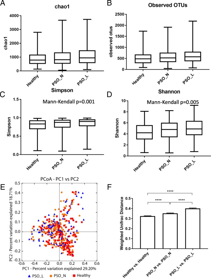
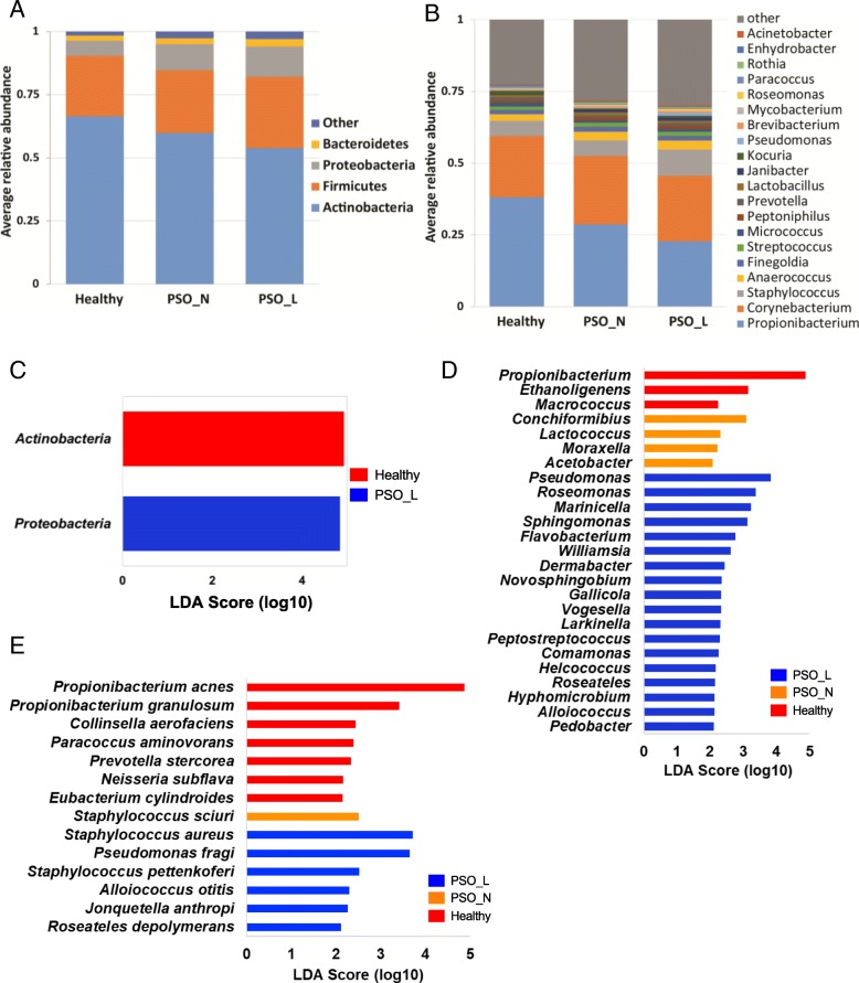
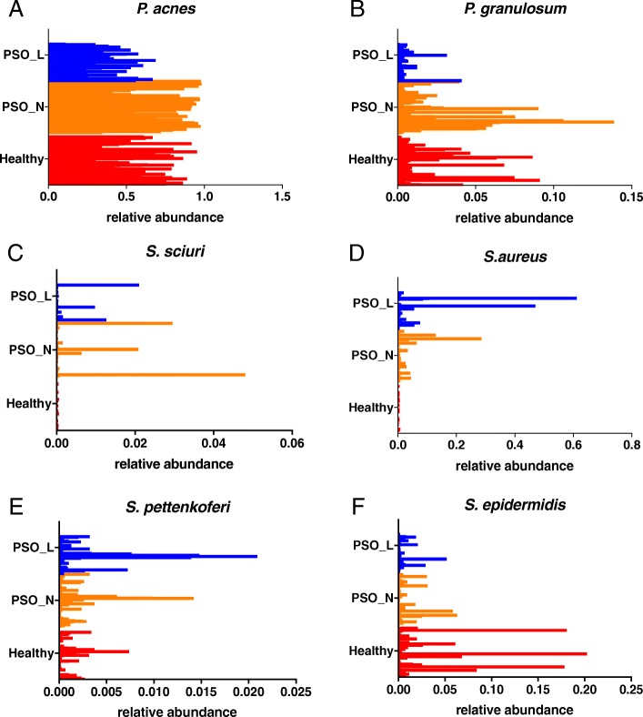
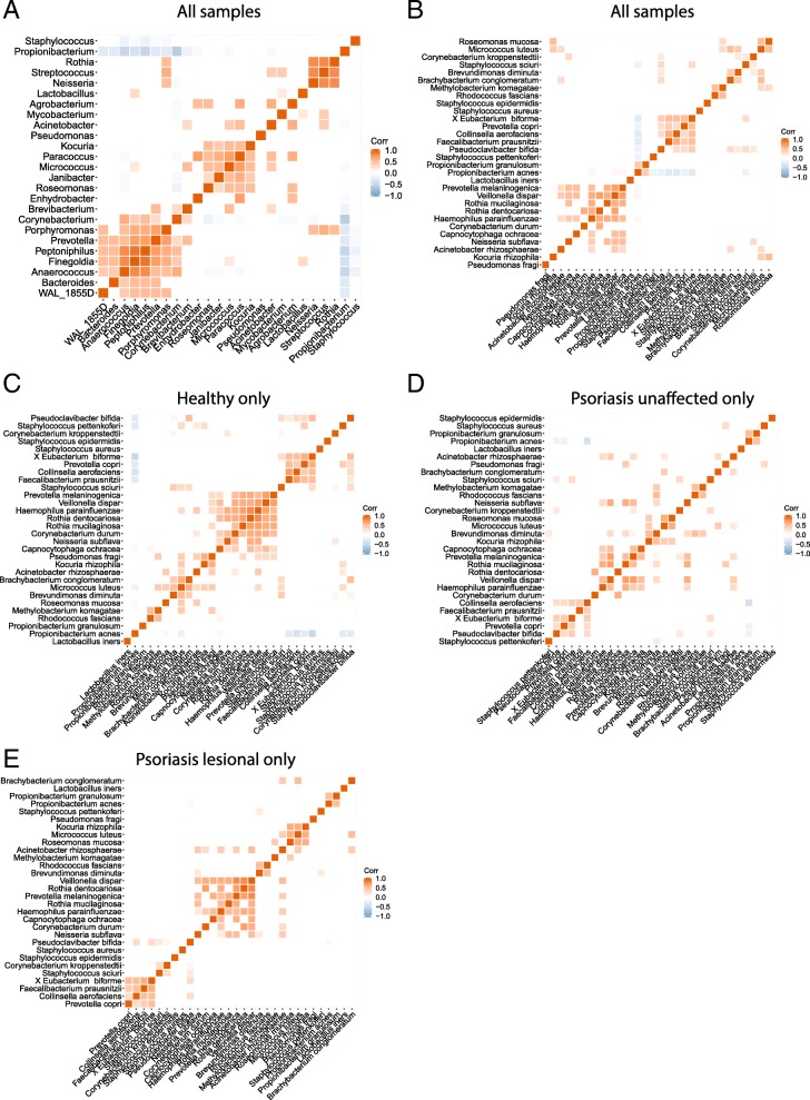
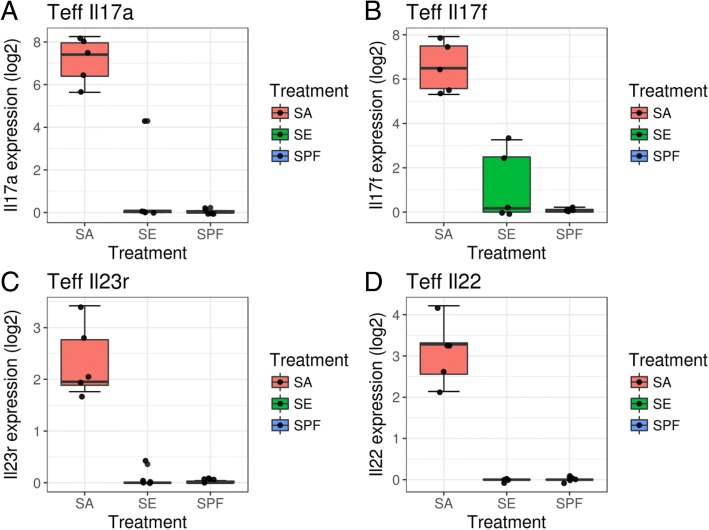
Results: Skin microbiome profiling based on sequencing the 16S rRNA V1-V3 variable region revealed significant differences between the psoriasis-associated and healthy skin microbiota. Comparing the overall community structures, psoriasis-associated microbiota displayed higher diversity and more heterogeneity compared to healthy skin bacterial communities. Specific microbial signatures were associated with psoriatic lesional, psoriatic non-lesional, and healthy skin. Specifically, relative enrichment of Staphylococcus aureus was strongly associated with both lesional and non-lesional psoriatic skin. In contrast, Staphylococcus epidermidis and Propionibacterium acnes were underrepresented in psoriatic lesions compared to healthy skin, especially on the arm, gluteal fold, and trunk. Employing a mouse model to further study the impact of cutaneous Staphylcoccus species on the skin T cell differentiation, we found that newborn mice colonized with Staphylococcus aureus demonstrated strong Th17 polarization, whereas mice colonized with Staphylococcus epidermidis or un-colonized controls showed no such response.

Conclusion: Our results suggest that microbial communities on psoriatic skin is substantially different from those on healthy skin. The psoriatic skin microbiome has increased diversity and reduced stability compared to the healthy skin microbiome. The loss of community stability and decrease in immunoregulatory bacteria such as Staphylococcus epidermidis and Propionibacterium acnes may lead to higher colonization with pathogens such as Staphylococcus aureus, which could exacerbate cutaneous inflammation along the Th17 axis.


Conflict of interest statement

Competing interests

The authors declare that they have no competing interests.

Publisher’s Note

Springer Nature remains neutral with regard to jurisdictional claims in published maps and institutional affiliations.

Figures

Fig. 1
Fig. 1
Bacterial community diversity in healthy and psoriasis skin. Alpha diversity measured according to a chao1 index, b observed OTUs, c Simpson’s diversity index, and d Shannon index of healthy skin samples, psoriasis non-lesional samples, and psoriasis lesional skin samples. Significant trends of alpha diversity are identified by a Mann-Kendall trend test with p-value shown. e Principal coordinate analysis (PCoA) of the microbial community structures based on weighted UniFrac distance matrix for the first two principal axes. Each point on the PCoA plot represents a skin microbiome sample (red square = healthy, blue triangle = psoriasis lesional, and orange circle = psoriasis unaffected). The first principal coordinate explains 29.6% of variation, and the second principal coordinate explains 18.70% of the variation. f The average weighted UniFrac distances among samples within each disease state are shown in the box plot. The samples in the psoriatic lesional group are more heterogeneous than samples from healthy or psoriasis unaffected groups (one-way ANOVA with Tukey correction, ****p value < 0.0001)
Fig. 2
Fig. 2
Taxonomical compositions and microbial signatures associated with each disease state. a Phylum and b genus level compositions of skin microbiome in healthy skin (Healthy), psoriasis unaffected skin (PSO_N), and psoriatic lesional skin (PSO_L). Only the predominant taxa are shown. Other represents lower abundant taxa that are not plotted. Bacterial taxa that are enriched in samples from healthy skin (red), psoriatic lesional skin (blue: PSO_L), and psoriatic unaffected skin (orange: PSO_N) at c phylum, d genus, and e species level. No phyla were significantly enriched in psoriasis unaffected skin
Fig. 3
Fig. 3
Relative abundance of bacterial species in each disease state. Histograms represent the relative abundances of specific bacterial species in samples from healthy skin (red bars: Healthy), psoriatic lesions (blue bars: PSO_L), and psoriatic unaffected skin (orange bars: PSO_N). Samples from healthy skin and psoriatic unaffected skin are more abundant in a Propionibacterium acnes (p value = 0.0002; LDA effect size = 4.87) and b Propionibacterium granulosum (p value = 0.014; LDA effect size = 3.41). Samples from psoriatic skin (both unaffected and lesional) are more abundant in c Staphylococcus sciuri (p value = 0.032; LDA effect size = 2.51), d Staphylococcus aureus (p value = 0.007; LDA effect size = 3.72), and e Staphylococcus pettenkoferi (p value = 0.012; LDA effect size = 2.52). On the contrary, f Staphylococcus epidermidis shows a trend of increased abundance in healthy skin but the difference did not reach the statistical significance
Fig. 4
Fig. 4
Correlations between the most abundant bacterial genera and species. Correlation plots show the Spearman correlations among a the top 25 most abundant genera or b the top 30 most abundant species in all samples. Correlations among the top 30 most abundant bacterial species associated with c Healthy skin samples, d psoriasis unaffected samples, and e psoriasis lesional samples. Only the correlations with statistical significance are shown. Color and intensity indicate directions and strength of the correlation
Fig. 5
Fig. 5
Staphylococcus aureus exposure triggers Th17 response in effector T cells. mRNA expression (log2FPKM) of cutaneous effector T cells from specific pathogen-free (SPF) mice colonized with Staphylococcus aureus (SA), Staphylococcus epidermidis (SE), or none (SPF). Compared to the SPF control, the Stapylococcus aureus colonization triggers gene expression in a IL-17A (adj p value = 3.51e−7), b IL-17F (adj p value = 3.08e−6), c IL-23R (adj p value = 3.74e−8), and d IL-22 (adj p value = 1.01e09). Colonization with Staphylococcus epidermidis does not trigger Th17 response

References

    1. Roberson EDO, Bowcock AM. Psoriasis genetics: breaking the barrier. Trends Genet. 2010;26:415–423. doi: 10.1016/j.tig.2010.06.006. - DOI - PMC - PubMed
    1. Afifi L, Danesh MJ, Lee KM, Beroukhim K, Farahnik B, Ahn RS, et al. Dietary behaviors in psoriasis: patient-reported outcomes from a U.S. national survey. Dermatol Ther (Heidelb) 2017;7:227–242. doi: 10.1007/s13555-017-0183-4. - DOI - PMC - PubMed
    1. Cai Y, Fleming C, Yan J. New insights of T cells in the pathogenesis of psoriasis. Cell Mol Immunol. 2012;9:302–309. doi: 10.1038/cmi.2012.15. - DOI - PMC - PubMed
    1. Grice EA, Segre JA. The skin microbiome. Nat Rev Microbiol. 2011;9:244–253. doi: 10.1038/nrmicro2537. - DOI - PMC - PubMed
    1. Belkaid Y, Tamoutounour S. The influence of skin microorganisms on cutaneous immunity. Nat Rev Immunol. 2016;16:353–366. doi: 10.1038/nri.2016.48. - DOI - PubMed

Publication types