. 2018 Sep 5;11(1):113.
doi: 10.1186/s13045-018-0656-7.

The lncRNA NEAT1 activates Wnt/β-catenin signaling and promotes colorectal cancer progression via interacting with DDX5

Affiliations

The lncRNA NEAT1 activates Wnt/β-catenin signaling and promotes colorectal cancer progression via interacting with DDX5

Meng Zhang et al. J Hematol Oncol. .

Abstract

Background: The long noncoding RNA nuclear-enriched abundant transcript 1 (NEAT1) has been reported to be overexpressed in colorectal cancer (CRC). However, its underlying mechanisms in the progression of CRC have not been well studied.

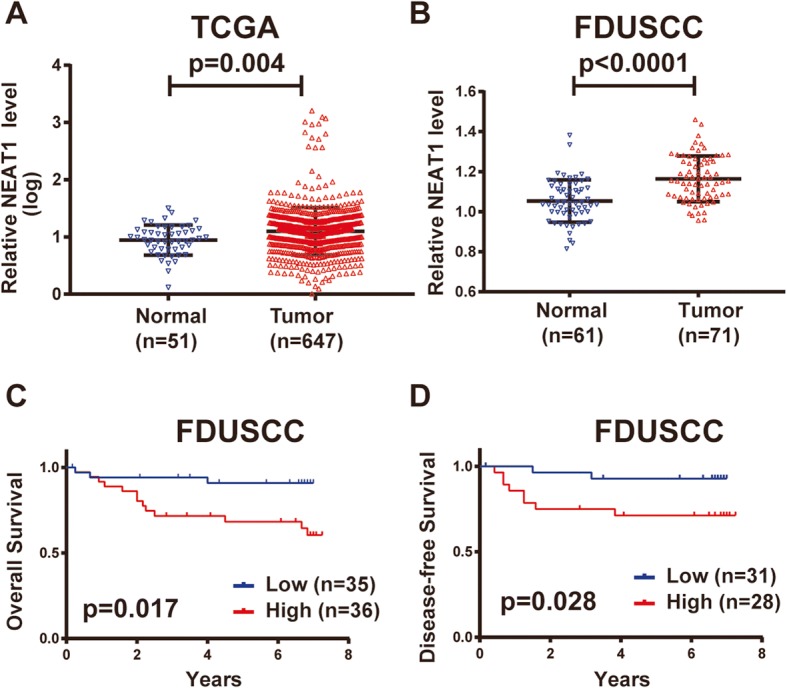
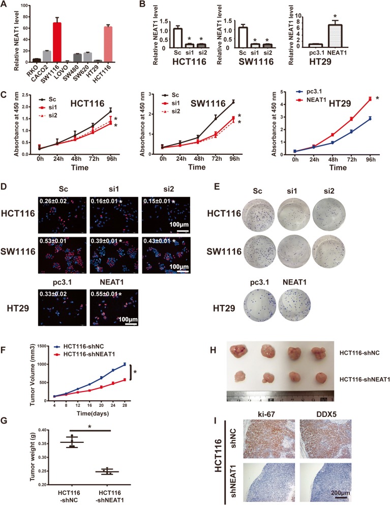
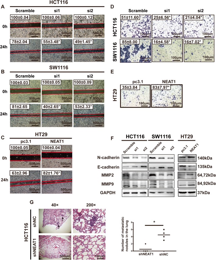
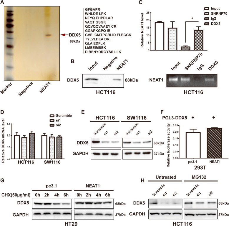
Methods: To investigate the clinical significance of NEAT1, we analyzed its expression levels in a publicly available dataset and in 71 CRC samples from Fudan University Shanghai Cancer Center. Functional assays, including the CCK8, EdU, colony formation, wound healing, and Transwell assays, were used to determine the oncogenic role of NEAT1 in human CRC progression. Furthermore, RNA pull-down, mass spectrometry, RNA immunoprecipitation, and Dual-Luciferase Reporter Assays were used to determine the mechanism of NEAT1 in CRC progression. Animal experiments were used to determine the role of NEAT1 in CRC tumorigenicity and metastasis in vivo.

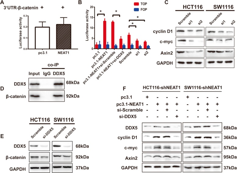
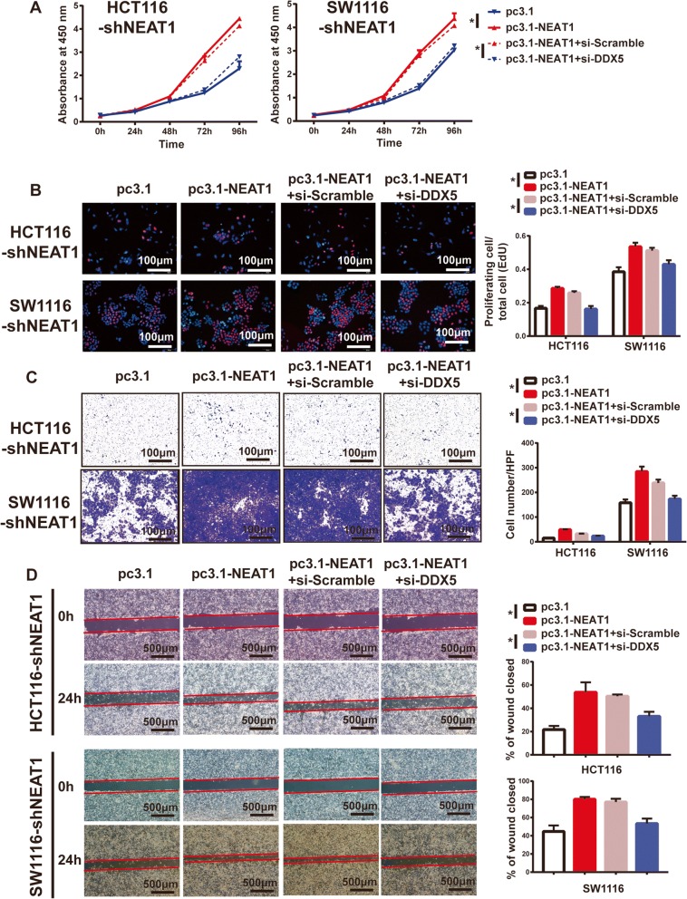
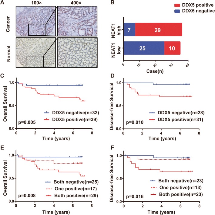
Results: NEAT1 expression was significantly upregulated in CRC tissues compared with its expression in normal tissues. Altered NEAT1 expression led to marked changes in proliferation, migration, and invasion of CRC cells both in vitro and in vivo. Mechanistically, we found that NEAT1 directly bound to the DDX5 protein, regulated its stability, and sequentially activated Wnt signaling. Our study showed that NEAT1 indirectly activated the Wnt/β-catenin signaling pathway via DDX5 and fulfilled its oncogenic functions in a DDX5-mediated manner. Clinically, concomitant NEAT1 and DDX5 protein levels negatively correlated with the overall survival and disease-free survival of CRC patients.

Conclusions: Our findings indicated that NEAT1 activated Wnt signaling to promote colorectal cancer progression and metastasis. The NEAT1/DDX5/Wnt/β-catenin axis could be a potential therapeutic target of pharmacological strategies.

Keywords: Colorectal cancer; DDX5; NEAT1; lncRNA; β-Catenin.


Conflict of interest statement

Ethics approval

The research protocol was reviewed and approved by the Ethics Committee of FDUSCC, and informed consent was obtained from all participants included in the study in agreement with institutional guidelines. This study complied with the Animal Care guidelines of FDUSCC.

Consent for publication

Informed consent for publication was obtained from all participants.

Competing interests

The authors declare that they have no competing interests.

Publisher’s Note

Springer Nature remains neutral with regard to jurisdictional claims in published maps and institutional affiliations.

Figures

Fig. 1
Fig. 1
NEAT1 is upregulated in human CRC patients and predicts a poor prognosis. a NEAT1 expression in TCGA CRC RNA-seq dataset (normal n = 51, tumor n = 647). b NEAT1 expression in the FDUSCC dataset (normal n = 61, tumor n = 71). cd Kaplan-Meier analyses of the FDUSCC dataset. c High NEAT1 expression predicted shorter OS than that of patients with low NEAT1 expression. d High NEAT1 expression predicted shorter DFS than that of patients with low NEAT1 expression
Fig. 2
Fig. 2
NEAT1 promoted CRC cell proliferation in vitro and in vivo. a The baseline NEAT1 RNA levels in eight CRC cell lines detected by RT-qPCR. b The efficiency of NEAT1 knockdown or overexpression was detected by RT-qPCR in the indicated cells transfected with siRNAs or plasmids (*p < 0.05). c CCK8 and d EdU assays showing that knockdown of NEAT1 suppressed cell proliferation in the HCT116 and SW1116 cell lines (*p < 0.05) and that upregulation of NEAT1 promoted cell proliferation in the HT29 cell line (*p < 0.05). e Colony formation assays for the indicated cells after transfection with siRNAs or plasmids (*p < 0.05). The nude mouse xenograft model showed that knockdown of NEAT1 decreased tumor growth (f) and the tumor weights (g) compared with those of the HCT116-shNC cells (*p < 0.05). h Representative images of tumors in nude mice. i Representative images of IHC staining for ki-67 and DDX5 (Sc represents scramble)
Fig. 3
Fig. 3
Repression of NEAT1-inhibited cell invasion and migration in vitro and in vivo. Representative images (× 40) of wound healing assays in HCT116 (a), SW1116 (b), and HT29 (c) cells (*p < 0.05). d Representative images (× 200) of Transwell invasion assays for the indicated cells (*p < 0.05). e Representative images of Transwell invasion assays for HT29 cells. f Western blotting results for N-cadherin, E-cadherin, MMP2, and MMP9. g Representative images of lung metastasis in nude mice with HE staining
Fig. 4
Fig. 4
NEAT1 binds to the DDX5 protein and enhances its stability. a RNA pull-down assay after silver staining and b western blotting to detect DDX5 protein expression in HCT116 cells. c RIP assay showing that DDX5 interacted with NEAT1 in HCT116 cells. The RT-qPCR products were analyzed by electrophoresis (below) (*p < 0.05). d The DDDX5 mRNA and e protein levels after transfection of the cells with siRNAs (*p < 0.05). f Dual-Luciferase Assays to assess DDX5 promoter activity. g Western blotting showing the DDX5 protein level after treatment of the cells with CHX (50 μg/mL) (*p < 0.05). h Western blotting showing the DDX5 protein level after treatment of the cells with or without MG132 (10 μmol/mL)
Fig. 5
Fig. 5
The influence of NEAT1 on β-catenin activation was dependent on DDX5. Dual-Luciferase Assays for β-catenin 3′-UTR (a) and TOP/FOP activity (b). c The Axin2, cyclin D1, and c-myc protein levels in the indicated cells (*p < 0.05). d Co-IP to detect the interaction of endogenous DDX5 and β-catenin in HCT116 cells. e Knockdown of DDX5 reduced β-catenin expression. f The DDX5, Axin2, cyclin D1, and c-myc protein levels after rescue of NEAT1 expression in shNEAT1 stable cells with or without si-DDX5
Fig. 6
Fig. 6
NEAT1 facilitated CRC cell progression in a DDX5-mediated manner. a CCK-8 and b EdU assay results showing that knockdown of DDX5 partially attenuated the enhanced cell proliferation induced by overexpression of NEAT1 in HCT116-shNEAT1 and SW1116-shNEAT1 cells (*p < 0.05). Representative images of the c Transwell invasion assays and d wound healing assays showing that DDX5 repression rescued the enhanced invasion and migration abilities induced by NEAT1 overexpression (*p < 0.05)
Fig. 7
Fig. 7
NEAT1 and DDX5 expression in clinical CRC samples. a Representative images of DDX5 detected by IHC. b The chi-square test identified an association between DDX5 and NEAT1 in the CRC samples (n = 71, p < 0.01). cf Kaplan-Meier analyses of the FDUSCC dataset. Patients with high DDX5 expression had poorer OS (c) and DFS (d) than those with negative expression. The patients were divided into three groups based on NEAT1 and DDX5 expression (negative or positive). Both positive groups had the poorest prognoses with the lowest OS (e) and DFS (f)

References

    1. Siegel R, Desantis C, Jemal A. Colorectal cancer statistics, 2014. CA Cancer J Clin. 2014;64(2):104–117. doi: 10.3322/caac.21220. - DOI - PubMed
    1. Dow LE, O'Rourke KP, Simon J, Tschaharganeh DF, van Es JH, Clevers H, et al. Apc restoration promotes cellular differentiation and reestablishes crypt homeostasis in colorectal cancer. Cell. 2015;161(7):1539–1552. doi: 10.1016/j.cell.2015.05.033. - DOI - PMC - PubMed
    1. Kong J, Sun W, Li C, Wan L, Wang S, Wu Y, et al. Long non-coding RNA LINC01133 inhibits epithelial-mesenchymal transition and metastasis in colorectal cancer by interacting with SRSF6. Cancer Lett. 2016;380(2):476–484. doi: 10.1016/j.canlet.2016.07.015. - DOI - PubMed
    1. Lee JT. Epigenetic regulation by long noncoding RNAs. Science. 2012;338(6113):1435–1439. doi: 10.1126/science.1231776. - DOI - PubMed
    1. Huang X, Xiao R, Pan S, Yang X, Yuan W, Tu Z, et al. Uncovering the roles of long non-coding RNAs in cancer stem cells. J Hematol Oncol. 2017;10(1):62. doi: 10.1186/s13045-017-0428-9. - DOI - PMC - PubMed

Publication types