. 2018 Sep 5;51(1):33.
doi: 10.1186/s40659-018-0182-7.

Increased ROS production and DNA damage in monocytes are biomarkers of aging and atherosclerosis

Affiliations

Increased ROS production and DNA damage in monocytes are biomarkers of aging and atherosclerosis

Thais A Jacinto et al. Biol Res. .

Abstract

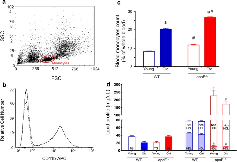
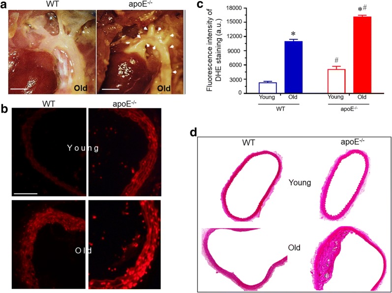
Background: New evidence demonstrates that aging and dyslipidemia are closely associated with oxidative stress, DNA damage and apoptosis in some cells and extravascular tissues. However, in monocytes, which are naturally involved in progression and/or resolution of plaque in atherosclerosis, this concurrence has not yet been fully investigated. In this study, we evaluated the influence of aging and hypercholesterolemia on serum pro-inflammatory cytokines, oxidative stress, DNA damage and apoptosis in monocytes from apolipoprotein E-deficient (apoE-/-) mice compared with age-matched wild-type C57BL/6 (WT) mice. Experiments were performed in young (2-months) and in old (18-months) male wild-type (WT) and apoE-/- mice.

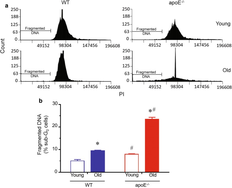
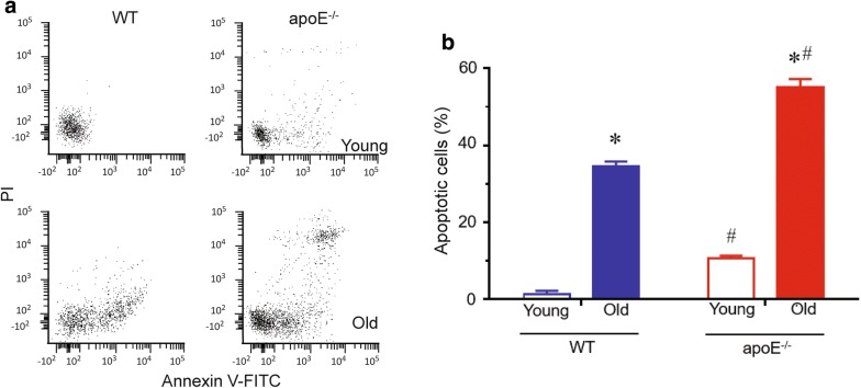
Results: Besides the expected differences in serum lipid profile and plaque formation, we observed that atherosclerotic mice exhibited a significant increase in monocytosis and in serum levels of pro-inflammatory cytokines compared to WT mice. Moreover, it was observed that the overproduction of ROS, led to an increased DNA fragmentation and, consequently, apoptosis in monocytes from normocholesterolemic old mice, which was aggravated in age-matched atherosclerotic mice.

Conclusions: In this study, we demonstrate that a pro-inflammatory systemic status is associated with an impairment of functionality of monocytes during aging and that these parameters are fundamental extra-arterial contributors to the aggravation of atherosclerosis. The present data open new avenues for the development of future strategies with the purpose of treating atherosclerosis.

Keywords: Apoptosis; Atherosclerosis; Proinflammatory cytokines; apoE knockout mice.


Figures

Fig. 1
Fig. 1
a The FSC scatter data provide information on the relative size of the cells, whereas the SSC data estimate the granularity. Dot plot where each dot represents one event. Here only 10,000 events are shown to avoid oversaturation of dots. Monocyte population defined by FSC and SSC properties are shown on the graph. b Peripheral blood cells were stained with anti-CD11b-APC antibody or isotype control to confirm the monocyte population. The flow cytometric immunophenotipic data revealed that the subset of monocytes defined by FSC and SSC expressed CD11b antigen (~ 40% positive cells). c Monocytes as percentage of whole lysed (depleted erythrocytes) peripheral blood cells. Bars graph represents the proportion of total circulating monocytes in apoE−/− and age-matched WT mice. d Lipid profile in apoE−/− and WT mice. Values are means ± SEM, *p < 0.05 vs. the respective young group, #p < 0.05 vs. age-matched WT group
Fig. 2
Fig. 2
Serum cytokine profile. Protein levels of pro-inflammatory cytokines in apoE−/− versus WT mice. Serum levels of IL-6 (a), TNFα (b), MCP-1 (c), INF-γ (d), IL-10 (e) and IL-12 (f) were evaluated by flow cytometry. Atherosclerotic aged mice showed augmented pro-inflammatory cytokine expression compared with WT. Values are means ± SEM, #p < 0.05 vs. age-matched WT group
Fig. 3
Fig. 3
Increased systemic ROS production in apoE−/− mice. a Top panel shows representative histogram with DHE staining of monocytes. b Bar graph shows the difference on MFI of DHE measured by flow cytometry in monocytes. Blue line: old-WT; red line: old-apoE−/−. Values are means ± SEM, *p < 0.05 vs. the respective young group, #p < 0.05 vs. age-matched WT group
Fig. 4
Fig. 4
a Representative photographs of mice aortic arch anatomy in situ. Atherosclerotic plaque formation is notably greater in apoE−/− mice, while no lesion was present in the age-matched WT control. Arrow heads indicates plaque deposition (4×; Bar = 2 mm). b DHE staining of representative aortic arch cross-sections. Atherosclerotic mice showed increased DHE-positive cells compared to WT (magnification 400×; Bar = 100 µm). c Quantification of ROS production. Aging increased ROS production in aortic arch cross-section and it was aggravated through hypercholesterolemia. d Representative micrographs of aortic arch stained with hematoxylin and eosin. Values are means ± SEM, **p < 0.01 vs. WT. *p < 0.05 vs. the respective young group, #p < 0.05 vs. age-matched WT group
Fig. 5
Fig. 5
DNA content analysis. a Typical histograms of cell cycle distribution of monocytes from young and old WT and apoE−/− mice. b Bar graph shows percentage of cells with fragmented DNA in monocytes. Values are mean ± SEM. *p < 0.05 vs. the respective young group, #p < 0.05 vs. age-matched WT group
Fig. 6
Fig. 6
Flow cytometric determination of apoptosis. a Representative dot-plots of Annexin VFITC and PI dual color flow cytometry for monocytes. b Bar graph shows average percentage of apoptotic cells (Q2 + Q4) from monocytes of WT and apoE−/− mice. Values are mean ± SEM. *p < 0.05 vs. the respective young group, #p < 0.05 vs. age-matched WT group

References

    1. Rudolf J, Lewandrowski KB. Cholesterol, lipoproteins, high-sensitivity c-reactive protein, and other risk factors for atherosclerosis. Clin Lab Med. 2014 - PubMed
    1. Libby P, Bornfeldt KE, Tall AR. Atherosclerosis: successes, surprises, and future challenges. Circ Res. 2016;118(4):531–534. doi: 10.1161/CIRCRESAHA.116.308334. - DOI - PMC - PubMed
    1. Herrington W, Lacey B, Sherliker P, Armitage J, Lewington S. Epidemiology of atherosclerosis and the potential to reduce the global burden of atherothrombotic disease. Circ Res. 2016;118(4):535–546. doi: 10.1161/CIRCRESAHA.115.307611. - DOI - PubMed
    1. Geng YJ, Jonasson L. Linking immunity to atherosclerosis: implications for vascular pharmacology-a tribute to Göran K. Hansson. Vasc Pharmacol. 2012 - PMC - PubMed
    1. Libby P, Ridker PM, Hansson GK. Leducq transatlantic network on atherothrombosis. Inflammation in atherosclerosis: from pathophysiology to practice. J Am Coll Cardiol. 2009 - PMC - PubMed