. 2018 Nov;210(3):907-923.
doi: 10.1534/genetics.118.301518. Epub 2018 Sep 5.

Multiple Histone Methyl-Lysine Readers Ensure Robust Development and Germline Immortality in Caenorhabditis elegans

Affiliations

Multiple Histone Methyl-Lysine Readers Ensure Robust Development and Germline Immortality in Caenorhabditis elegans

Arneet L Saltzman et al. Genetics. 2018 Nov.

Abstract

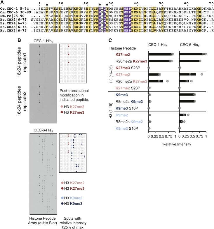
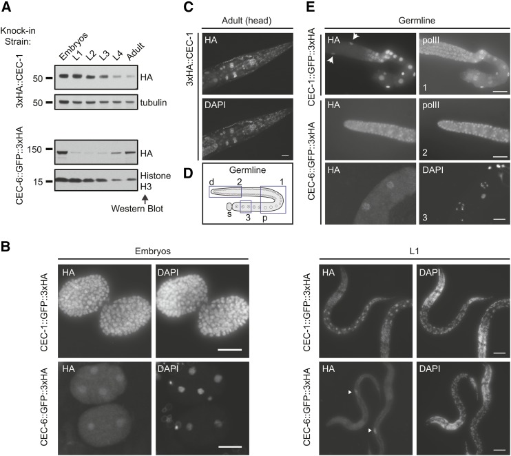
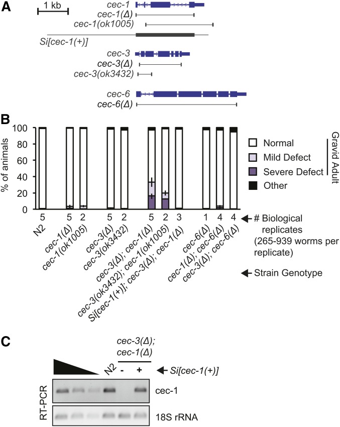
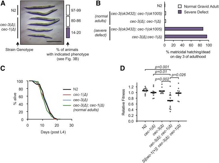
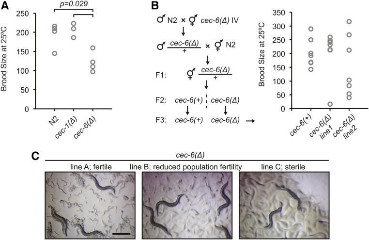
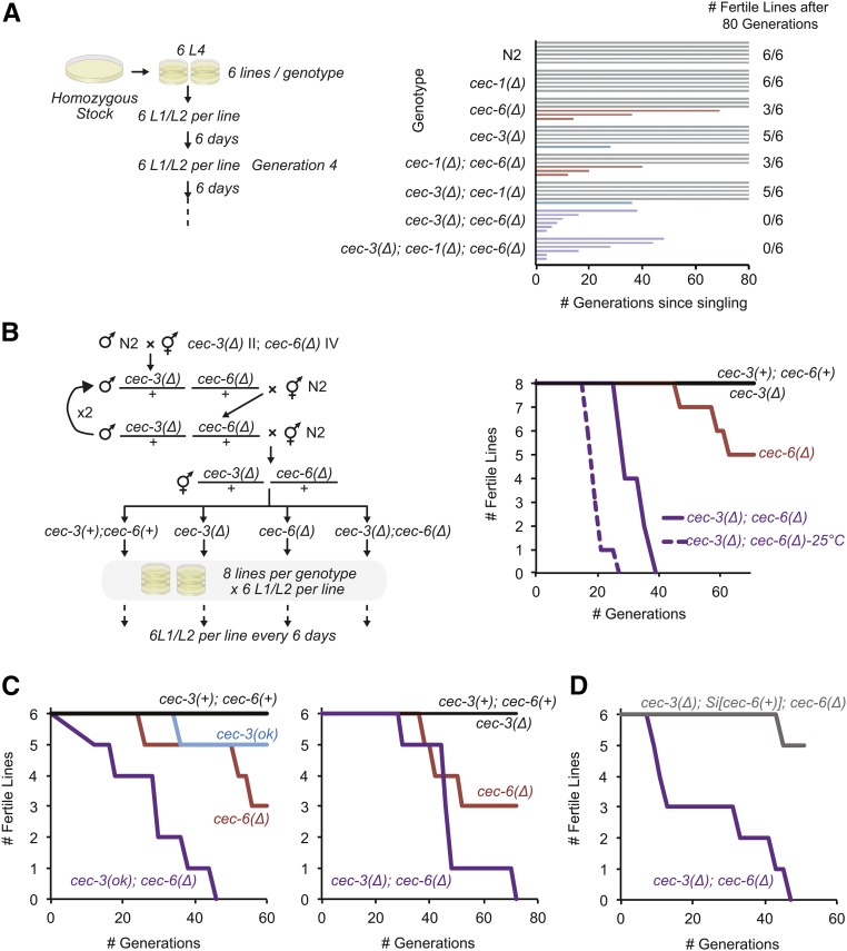
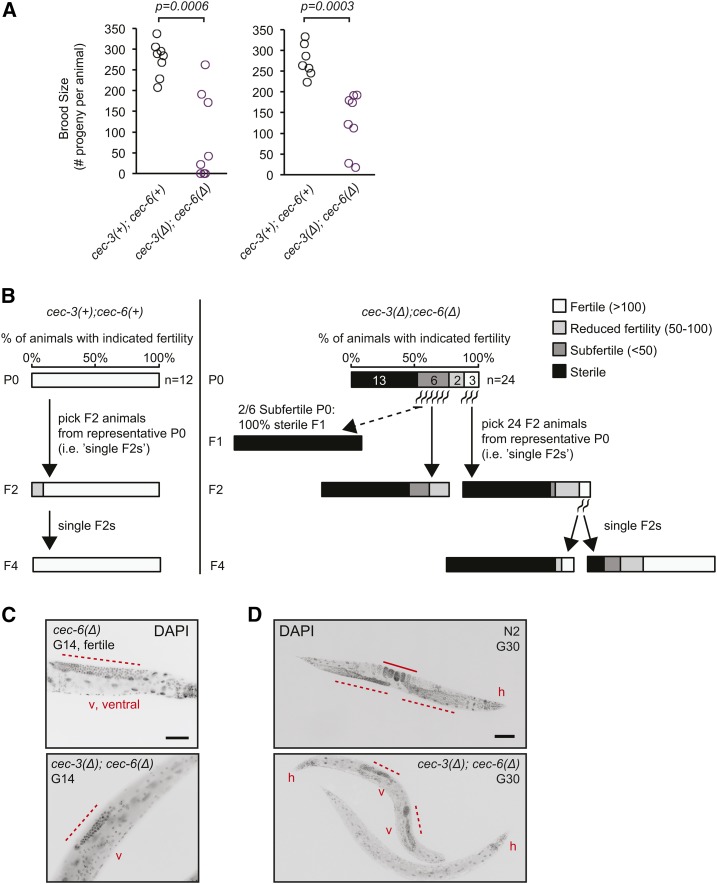
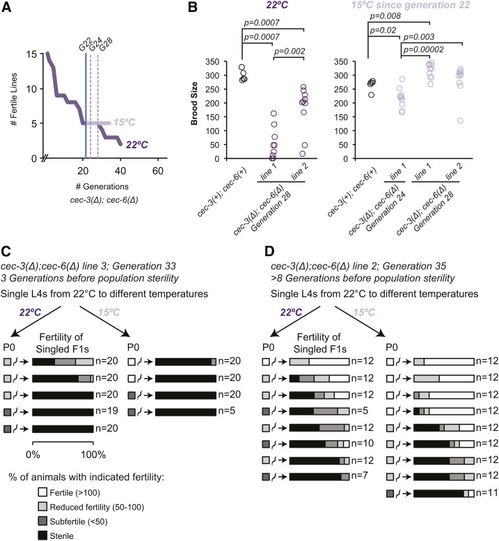
Chromatin modifications, including methylation of histone H3 at lysine 27 (H3K27me) by the Polycomb group proteins, play a broadly conserved role in the maintenance of cell fate. Diverse chromatin organization modifier (chromo) domain proteins act as "readers" of histone methylation states. However, understanding the functional relationships among chromo domains and their roles in the inheritance of gene expression patterns remains challenging. Here, we identify two chromo-domain proteins, CEC-1 and CEC-6, as potential readers of H3K27me in Caenorhabditis elegans, where they have divergent expression patterns and contribute to distinct phenotypes. Both cec-1 and cec-6 genetically interact with another chromo-domain gene, cec-3, a reader of H3K9 methylation. Combined loss of cec-1 and cec-3 leads to developmental defects in the adult that result in decreased fitness. Furthermore, loss of cec-6 and cec-3 surprisingly leads to a progressive loss of fertility across generations, a "mortal germline" phenotype. Our results provide evidence of functional compensation between H3K27me and H3K9me heterochromatin pathways, and show that histone methylation readers contribute to both somatic development and transgenerational fitness.

Keywords: C. elegans; CEC; H3K27me3; PRC2; Polycomb proteins; chromodomain proteins; gene silencing; histone methyl-lysine readers; mortal germline; worm.


Figures

Figure 1
Figure 1
Binding specificity of C. elegans chromo-domain proteins CEC-1 and CEC-6 for histone H3 peptides with tri- or dimethylation at lysine (K) 27 or K9 (H3K27me/H3K9me). (A) Multiple sequence alignment showing amino acid similarity of the chromo domains of CEC-1 and CEC-6 to those of Drosophila polycomb (Dm_Pc) and the human Chromobox (Hs_CBX) proteins. Conserved aromatic residues involved in methyl-lysine binding (Fischle et al. 2003; Min et al. 2003) are indicated by asterisks. Residue coloring: purple boxes, amino acids identical in all seven sequences; yellow boxes and bold text, amino acids with similar properties in all seven sequences; yellow boxes, bold and normal text, identical amino acids in six of seven sequences. (B) Binding of recombinant His6-tagged proteins to histone peptide arrays detected by HRPase-conjugated anti-His antibody and chemiluminescence. Arrays contain 384 peptide spots in duplicates of 19mer histone peptides bearing single or multiple post-translational modifications. The map highlights all spots with a relative intensity ≥ 0.25, where the array spot with the greatest intensity is set to 1, as described in the Materials and Methods. Colors show that the indicated spots contain H3K27me3/2 or H3K9me3/2. (C) Quantification of peptide array signals showing relative binding of CEC-1 or CEC-6 to H3K27me3/2 and H3K9me3/2 in combination with other peptide modifications. R8/26me2s/a refers to symmetric or asymmetric arginine dimethylation. Relative intensities of individual peptide spots are indicated by circles and the average is shown as a bar. For additional quantification, see also Figure S2 and Table S2.
Figure 2
Figure 2
Distinct expression patterns of C. elegans chromo-domain proteins CEC-1 and CEC-6. (A) Relative expression levels of CEC-1 and CEC-6 across developmental stages. The 3xHA (hemagglutinin) epitope was inserted in-frame at the endogenous locus using clustered regularly interspaced short palindromic repeats/Cas9 genome editing (see also Figure S3C). L1–L4, larval stages 1–4. (B) Localization of CEC-1 and CEC-6 by anti-HA tag immunofluorescence in embryos and L1 animals. The arrowheads [(B), lower right] indicate the primordial germ cells (see also Figure S3D). (C) Broad nuclear expression of CEC-1 in the adult head. (D) Schematic of an isolated germline arm indicating three regions depicted in (E). The distal germline is a syncytium, with germ cells surrounding a central canal. d, distal; p, proximal; and s, spermatheca. (E) Expression of CEC-1 and CEC-6 in dissected germlines. The arrows (top panel) indicate nuclei of the somatic gonad cells. Germlines in (C) were costained with an antibody detecting the RNA polymerase C-terminal domain (polII) to control for antibody penetration.
Figure 3
Figure 3
Loss of cec-1 and cec-3 leads to adult-onset defects with partial penetrance. (A) Schematic of alleles and transgenes used in this study (see also Figure S5). The borders of deletion alleles are indicated below the gene diagrams. The genomic fragment used for the Minimal Mos1 (miniMos) transgene rescue (Si[cec-1(+)]) is indicated. (B) Quantification of abnormal gross morphology phenotypes in synchronized worm populations of indicated genotypes grown to gravid adulthood. Error bars represent the range of proportions of abnormal worms from all biological replicates scored. For images of abnormal adult animals, see Figure 4A. (C) The miniMos cec-1 rescue transgene (Si[cec-1(+)]) mRNA is expressed at similar levels to endogenous cec-1, as assayed by RT-PCR.
Figure 4
Figure 4
Synthetic fitness defect in cec-3Δ;cec-1Δ adult animals. (A) Stereoscope image of wild-type (N2) and age-matched cec-3Δ;cec-1Δ normal or severely abnormal gravid animals as quantified in Figure 3B. Bar, 0.5 mm. (B) Nearly all severely abnormal cec-3Δ;cec-1Δ adults do not survive past day 3 of adulthood. (C) The life span of phenotypically normal cec-3Δ;cec-1Δ animals is not significantly different from wild-type or single-mutant animals. Number of animals scored: N2, 55; cec-1Δ, 53; cec-3Δ, 58; and cec-3Δ;cec-1Δ, 55. (D) Phenotype of cec-3Δ;cec-1Δ animals in a competitive fitness assay. Animals of the indicated genotypes were grown in competition with a GFP-marked strain, cec-1(Pmyo-2::GFP+). The P-values indicate a two-sample Student’s t-test.
Figure 5
Figure 5
Variability in fertility of cec-6Δ animals. (A) Brood size per animal for worms of indicated genotypes. The distribution of the cec-6Δ animals is shifted relative to the N2 and cec-1Δ animals (Wilcoxon rank-sum test, one-sided). The distributions of N2 and cec-1Δ brood sizes were not significantly different. (B) Brood size per animal after outcrossing to N2 as indicated in the scheme. Differences in distributions were not significant (Wilcoxon rank-sum test, two-sided). (C) Stereoscope images of growth plates 6 days after six L1 worms were plated. Images represent lines at various stages of population fertility. When lines became sterile, most animals had a dark appearance with a clear area where embryos are normally located (Capowski et al. 1991; Katz et al. 2009) (right panel). Bar, 0.5 mm.
Figure 6
Figure 6
Germline immortality is compromised in cec-3Δ;cec-6Δ animals. (A and B) Mortal germline assays starting from homozygous mutant (A) or freshly outcrossed (B) strains. In (B), strains harboring cec-3Δ and cec-6Δ deletions were backcrossed to N2 (wild-type) worms four times consecutively, while maintaining both cec-3 and cec-6 in a heterozygous state until the final generation. All assays were performed at 22° except as indicated [dotted line in (B)]. (C) Independent biological replicates of mortal germline assays. Animals with indicated alleles were outcrossed five times to N2 before the start of the assays. Experiments in the left and right panels were conducted using independent alleles of cec-3 [left panel: cec-3(ok3432), from the C elegans knockout consortium; right panel: cec-3Δ, this study]. (D) The mortal germline of cec-3Δ;cec-6Δ animals is rescued by a cec-6(+) single-copy transgene inserted using Minimal Mos1 (see text).
Figure 7
Figure 7
Germline and fertility defects in individual late-generation cec-3Δ;cec-6Δ animals. (A) Fertility defects of late-generation cec-3Δ;cec-6Δ animals. Individual worms were randomly selected for brood size assays 14 (left) or 10 (right) generations (G) before population sterility, corresponding to G58 and G62 from the mortal germline assay in Figure 6C, right. The P-values represent a two-sided Wilcoxon rank-sum test. (B) Transgenerational fertility of late-generation single animals during the mortal germline assay. For cec-3Δ;cec-6Δ animals (right panel), F2 progeny were analyzed from P0 animals with varying levels of fertility (G66 from the mortal germline assay in Figure 6C, right). All assays were performed at 22°. (C and D) Germline morphology in fertile, sterile, or near-sterile DAPI-stained fixed animals of the indicated genotypes. Animals in (C and D) are from the mortal germline assays shown in Figure 6, A and B, respectively, at the indicated generations. Bar, (C) 50 μm; (D) 100 μm. v, ventral; h, head. Dotted lines indicate the distal germline and the solid line in (C) indicates embryos in the uterus. For additional images, see Figure S6.
Figure 8
Figure 8
Effect of temperature downshift on late-generation cec-3Δ;cec-6Δ animals. (A) A mortal germline assay was initiated using 15 lines at 22°. When five fertile lines remained [generation (G) 22, blue vertical line), lines were passaged to both 22 and 15° and mrt assays were continued in parallel at both temperatures. The break on the x-axis indicates that the generation count was restarted when these 15 lines were initiated from fertile plates from the mortal germline assay presented in Figure 6C (right panel). (B) Brood size assays were conducted on animals selected from plates at each temperature two and six generations (G24 and G28, dotted gray vertical lines) after the temperature shift. The P-values are shown for all pairwise comparisons where P < 0.05 (two-sided Wilcoxon rank-sum test). (C and D) L4 animals from the 22° mrt assay (A) were singled to the indicated temperatures. Their fertility was scored as indicated by the color scale and described in Materials and Methods. Approximately 12–20 F1 animals were singled per line and their fertility was also scored. Assays were initiated at the generation number indicated in the figure.

Similar articles

Cited by

References

    1. Aasland R., Stewart A. F., 1995. The chromo shadow domain, a second chromo domain in heterochromatin-binding protein 1, HP1. Nucleic Acids Res. 23: 3168–3173. 10.1093/nar/23.16.3168 - DOI - PMC - PubMed
    1. Agostoni E., Albertson D., Wittmann C., Hill F., Tobler H., et al. , 1996. cec-1, a soma-specific chromobox-containing gene in C. elegans. Dev. Biol. 178: 316–326. 10.1006/dbio.1996.0221 - DOI - PubMed
    1. Ahmed S., 2006. Uncoupling of pathways that promote postmitotic life span and apoptosis from replicative immortality of Caenorhabditis elegans germ cells. Aging Cell 5: 559–563. 10.1111/j.1474-9726.2006.00244.x - DOI - PubMed
    1. Ahmed S., Hodgkin J., 2000. MRT-2 checkpoint protein is required for germline immortality and telomere replication in C. elegans. Nature 403: 159–164. 10.1038/35003120 - DOI - PubMed
    1. Altschul S. F., Gish W., Miller W., Myers E. W., Lipman D. J., 1990. Basic local alignment search tool. J. Mol. Biol. 215: 403–410. 10.1016/S0022-2836(05)80360-2 - DOI - PubMed

Publication types