. 2018 Sep 5;3(5):e00400-18.
doi: 10.1128/mSphere.00400-18.

The Transcription Factor VdHapX Controls Iron Homeostasis and Is Crucial for Virulence in the Vascular Pathogen Verticillium dahliae

Affiliations

The Transcription Factor VdHapX Controls Iron Homeostasis and Is Crucial for Virulence in the Vascular Pathogen Verticillium dahliae

Yonglin Wang et al. mSphere. .

Abstract

Iron homeostasis is essential for full virulence and viability in many pathogenic fungi. Here, we showed that the bZip transcription factor VdHapX functions as a key regulator of iron homeostasis for adaptation to iron-depleted and iron-excess conditions and is required for full virulence in the vascular wilt fungus, Verticillium dahliae Deletion of VdHapX impaired mycelial growth and conidiation under both iron starvation and iron sufficiency. Furthermore, disruption of VdHapX led to decreased formation of the long-lived survival structures of V. dahliae, known as microsclerotia. Expression of genes involved in iron utilization pathways and siderophore biosynthesis was misregulated in the ΔVdHapX strain under the iron-depleted condition. Additionally, the ΔVdHapX strain exhibited increased sensitivity to high iron concentrations and H2O2, indicating that VdHapX also contributes to iron or H2O2 detoxification. The ΔVdHapX strain showed a strong reduction in virulence on smoke tree seedlings (Cotinus coggygria) and was delayed in its ability to penetrate plant epidermal tissue.IMPORTANCE This study demonstrated that VdHapX is a conserved protein that mediates adaptation to iron starvation and excesses, affects microsclerotium formation, and is crucial for virulence of V. dahliae.

Keywords: HapX; Verticillium dahliae; fungal virulence; iron homeostasis; vascular wilt.


Figures

FIG 1
FIG 1
Deletion of VdHpaX impairs hyphal growth and conidiation in Verticillium dahliae. (A) Colonies of the wild-type strain (XS11), the ΔVdHapX strain, and the complemented strain (ΔVdHapX/VdHapX) grown on potato dextrose agar (PDA) and minimal medium (MM) plates for 14 days at 25°C. The indicated strains were incubated on MM plates with 0.06 mM Fe3+ and 0.4 mM BPS, respectively. (B) Relative mycelial growth of the indicated strains on MM with or without iron. The data were obtained by measuring the diameter of fungal colonies and were normalized to the growth of XS11 on MM. (C) Vertical dissection of the colonies on MM plates in the presence or absence of iron. (D) Relative dry biomass of the respective strains grown in liquid MM with or without iron for 14 days at 25°C. (E) Conidial production of the strains after growth for 7 days on liquid MM with or without iron. The error bars represent standard deviations based on three independent replicates, and asterisks represent significant differences (P < 0.01). Bar, 1 cm.
FIG 2
FIG 2
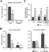
Disruption of VdHapX causes downregulation of iron-regulatory genes but not intracellular iron concentration in Verticillium dahliae. (A) Reverse transcription-quantitative PCR was utilized to determine transcript levels of VdHapX that were analyzed in XS11 and compared with those of the ΔVdHapX strain under iron-depleted MM (-Fe) or iron-replete MM (+Fe) conditions. The expression was normalized against the expression of the V. dahliae β-tubulin gene. Error bars indicate standard deviations from three independent experiments. (B) The expression of srbA and sreA was analyzed in XS11 and ΔVdHapX strains after 3 days in iron-depleted MM and transfer for 45 min to iron-depleted MM (-Fe) or iron-replete MM (+Fe). Both srbA expression and sreA expression were normalized against the V. dahliae β-tubulin gene. Values are the averages from four biological replicates, consisting of three technical replicates each. Error bars represent standard deviations. (C) Total intracellular iron concentration measurment. XS11, ΔVdHapX mutant, and ΔVdHapX/VdHapX complemented strains were grown in iron-depleted MM for 3 days and transferred for 45 min to iron-depleted MM (-Fe) or iron-replete MM (+Fe). The intracellular iron concentration was determined using an optical emission spectrometer. Error bars represent the standard deviations based on three independent replicates with three technical replicates. Asterisks represent significant differences (P < 0.01).
FIG 3
FIG 3
Genes involved in siderophore biosynthesis in Verticillium dahliae are misregulated in the ΔVdHapX strain during iron starvation. Reverse transcription-quantitative PCR analysis of postulated siderophore ferricrocin and ferrichrome C biosynthesis pathway genes based on information available in Aspergillus spp. (24). Transcript levels of sidA (VDAG_05313), sidC (VDAG_05314), sidD (VDAG_03964), and mirB (VDAG_07020) of V. dahliae wild-type XS11 and ΔVdHapX strains under iron starvation (-Fe) and iron-replete (+Fe) conditions. Data represent the means from three biological replicates. Averages of gene expression values were normalized against the β-tubulin gene. Error bars represent standard deviations. Asterisks represent significant differences: *, P < 0.05; **, P < 0.01.
FIG 4
FIG 4
The ΔVdHapX strain of Verticillium dahliae displays increased sensitivity to iron toxicity. (A) The wild-type XS11, ΔVdHapX, and ΔVdHapX/VdHapX complemented strains were grown on solid MM with specified iron availability (Fe2+ and Fe3+) conditions for 7 days at 25°C. Images of colony morphology were taken at 7 days. (B) Mycelial growth of the indicated strains grown as described for panel A. Data are representative of mean diameters of different colonies. Error bars represent the standard deviations based on three independent replicates with three technical replicates. (C and D) Expression of cccA encoding vacuolar iron transporter in XS11 and ΔVdHapX strains following normal iron conditions (0.03 mM Fe2+ or Fe3+) and high-iron conditions (10 mM Fe2+ or Fe3+) for 45 min. Averages of the gene expression values were normalized against the V. dahliae β-tubulin gene. Error bars represent standard deviations. The asterisks indicate a significant difference at P < 0.01.
FIG 5
FIG 5
Deletion of Verticillium dahliae VdHapX causes deregulation of genes involved in iron use. RT-qPCR analysis was performed in XS11 and ΔVdHapX strains grown in liquid MM for 3 days at 25°C and transferred for 45 min to iron-depleted MM (-Fe) or iron-replete MM (0.03 mM, +Fe). Transcript levels of acoA, lysF, hemA, cycA, VDAG_00564, and VDAG_04620 were analyzed under the different iron conditions shown. Averages of the gene expression values were normalized against the V. dahliae β-tubulin gene. Error bars represent standard deviations. Asterisks represent significant differences: *, P < 0.05; **, P < 0.01.
FIG 6
FIG 6
Deletion of VdHapX renders Verticillium dahliae more susceptible to H2O2. (A) Conidial suspensions (1 × 10⁵ spores) of each of the wild-type XS11, ΔVdHapX, and ΔVdHapX/VdHapX complemented strains were spread on potato dextrose agar plates. Sterile filter paper disks (5-mm diameter) were placed in the center of the plates, and 10 μl of an H2O2 solution (2.5%, 5%, and 10%) was added to each paper disk. The plates were incubated at 25°C for 4 days, and the inhibition zones were observed. (B) Bar chart of the inhibition zone of the above-described plates. Error bars represent the standard deviations based on three independent replicates. Asterisks indicate significant differences at P < 0.01. (C) Expression of genes involved in H2O2 detoxification in V. dahliae, such as cat1, sod, sod_Cu, and yap1. Strains were grown in minimal medium for 3 days and then transferred for 45 min to 1 mM H2O2. Averages of the gene expression values were normalized against the V. dahliae β-tubulin gene. Error bars represent standard deviations. Asterisks represent significant differences (P < 0.01).
FIG 7
FIG 7
Deletion of VdHapX affects melanized microsclerotium formation in Verticillium dahliae. (A) Colony morphology and microscopic examination of melanized microsclerotium formation of the XS11, ΔVdHapX, and ΔVdHapX/VdHapX complemented strains on a BM slide at 25°C for 4 days. Bars in the top, middle, and bottom panels represent 0.5 cm, 0.25 cm, and 20 mm, respectively. (B) Bar chart showing the relative ratio of microsclerotia produced by the indicated strains. Microsclerotia formed on slides coated with BM during a 7-day incubation period. Photographs of the microsclerotia in a field were taken and converted into an 8-bit grayscale image by using Image J. Using pixels as units in the measurement area, the pixel values of the covered area were measured and the relative ratio was calculated. There were three independent fields in which microsclerotia were enumerated. Error bars represent the standard deviations based on three independent replicates. The asterisk indicates a significant difference at P < 0.05.
FIG 8
FIG 8
Deletion of VdHapX compromises full virulence on host smoke trees and penetration into plant epidermal tissue. (A) One-year-old smoke tree seedlings were inoculated and incubated for 10 min with a 106-conidium/ml suspension of XS11, the ΔVdHapX strain, and the ΔVdHapX/VdHapX strain. The smoke tree seedlings were inoculated with distilled water (CK), also for a 10-min incubation period. All seedlings were replanted in soil for 35 days of growth, and the pictures were captured at 35 days after inoculation. (B) Colonies of each of the XS11, ΔVdHapX, and ΔVdHapX/VdHapX strains were grown on minimal medium overlaid with a cellophane membrane and incubated 7 dpi (top) and at 3 days after removal of the cellophane membrane (bottom). Bar, 1 cm. (C) Hyphopodia and penetration pegs formed on a cellophane membrane at 4 dpi. Bar, 10 µm. (D) Infection assays of onion epidermis examined at 24, 48, and 96 h postinoculation. Fungal hyphae were stained with trypan blue solution. Arrows indicate invasive hyphae (IH). Bars, 40 µm.

Similar articles

Cited by

References

    1. Lin H, Li L, Jia X, Ward DM, Kaplan J. 2011. Genetic and biochemical analysis of high iron toxicity in yeast: iron toxicity is due to the accumulation of cytosolic iron and occurs under both aerobic and anaerobic conditions. J Biol Chem 286:3851–3862. doi:10.1074/jbc.M110.190959. - DOI - PMC - PubMed
    1. Halliwell B, Gutteridge JM. 1984. Oxygen toxicity, oxygen radicals, transition metals and disease. Biochem J 219:1–14. doi:10.1042/bj2190001. - DOI - PMC - PubMed
    1. Haas H, Zadra I, Stoffler G, Angermayr K. 1999. The Aspergillus nidulans GATA factor SREA is involved in regulation of siderophore biosynthesis and control of iron uptake. J Biol Chem 274:4613–4619. doi:10.1074/jbc.274.8.4613. - DOI - PubMed
    1. Hortschansky P, Eisendle M, Al-Abdallah Q, Schmidt AD, Bergmann S, Thon M, Kniemeyer O, Abt B, Seeber B, Werner ER, Kato M, Brakhage AA, Haas H. 2007. Interaction of HapX with the CCAAT-binding complex—a novel mechanism of gene regulation by iron. EMBO J 26:3157–3168. doi:10.1038/sj.emboj.7601752. - DOI - PMC - PubMed
    1. Schrettl M, Beckmann N, Varga J, Heinekamp T, Jacobsen ID, Jochl C, Moussa TA, Wang S, Gsaller F, Blatzer M, Werner ER, Niermann WC, Brakhage AA, Haas H. 2010. HapX-mediated adaption to iron starvation is crucial for virulence of Aspergillus fumigatus. PLoS Pathog 6:e1001124. doi:10.1371/journal.ppat.1001124. - DOI - PMC - PubMed

Publication types

MeSH terms

LinkOut - more resources