. 2018 Sep 5;38(36):7822-7832.
doi: 10.1523/JNEUROSCI.3576-17.2018. Epub 2018 Aug 1.

Evaluating the Columnar Stability of Acoustic Processing in the Human Auditory Cortex

Affiliations

Evaluating the Columnar Stability of Acoustic Processing in the Human Auditory Cortex

Michelle Moerel et al. J Neurosci. .

Abstract

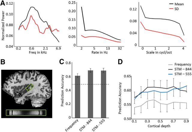
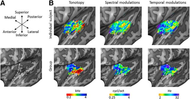
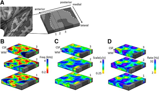
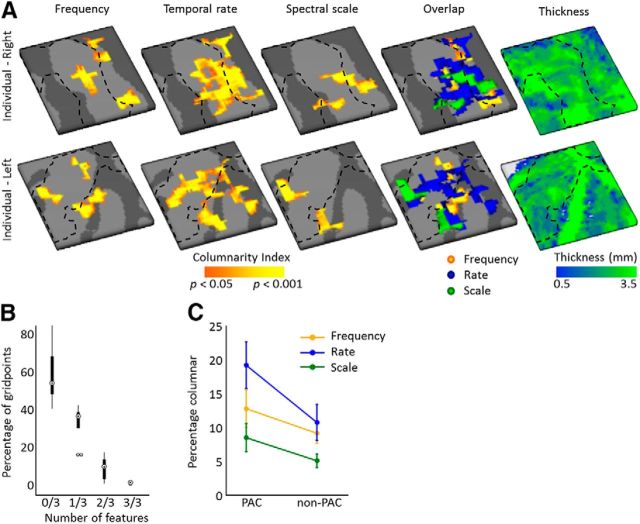
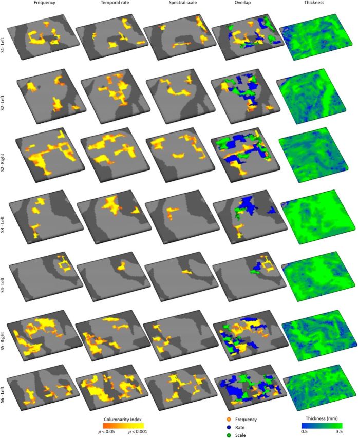
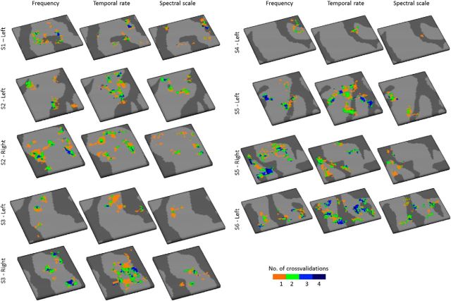
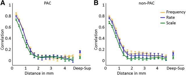
Using ultra-high field fMRI, we explored the cortical depth-dependent stability of acoustic feature preference in human auditory cortex. We collected responses from human auditory cortex (subjects from either sex) to a large number of natural sounds at submillimeter spatial resolution, and observed that these responses were well explained by a model that assumes neuronal population tuning to frequency-specific spectrotemporal modulations. We observed a relatively stable (columnar) tuning to frequency and temporal modulations. However, spectral modulation tuning was variable throughout the cortical depth. This difference in columnar stability between feature maps could not be explained by a difference in map smoothness, as the preference along the cortical sheet varied in a similar manner for the different feature maps. Furthermore, tuning to all three features was more columnar in primary than nonprimary auditory cortex. The observed overall lack of overlapping columnar regions across acoustic feature maps suggests, especially for primary auditory cortex, a coding strategy in which across cortical depths tuning to some features is kept stable, whereas tuning to other features systematically varies.SIGNIFICANCE STATEMENT In the human auditory cortex, sound aspects are processed in large-scale maps. Invasive animal studies show that an additional processing organization may be implemented orthogonal to the cortical sheet (i.e., in the columnar direction), but it is unknown whether observed organizational principles apply to the human auditory cortex. Combining ultra-high field fMRI with natural sounds, we explore the columnar organization of various sound aspects. Our results suggest that the human auditory cortex contains a modular coding strategy, where, for each module, several sound aspects act as an anchor along which computations are performed while the processing of another sound aspect undergoes a transformation. This strategy may serve to optimally represent the content of our complex acoustic natural environment.

Keywords: auditory cortex; columnar processing; spectrotemporal modulations; tonotopy; ultra-high field fMRI.


Figures

Figure 1.
Figure 1.
Sound characteristics, brain coverage, and performance of the encoding models. A, Mean and SD (in black and red, respectively) of the frequency, temporal modulation rate, and spectral modulation scale content of the 144 natural sounds is shown from left to right. B, The 3D GRASE acquisition covered HG, in addition to regions at its anterior and posterior adjacency. C, Performance of the models evaluated by the prediction accuracy of responses to test sounds (i.e., sound identification score) on the full dataset covering HG and regions at its anterior and posterior vicinity bilaterally (in 3 of 6 subjects) or on the left hemisphere (in 3 of 6 subjects). The performance of a frequency model is compared with that of two frequency-specific STM models, either with dimensions [8 frequencies × 4 spectral scales × 4 temporal rates] (STM-844), or with dimensions [5 frequencies × 5 spectral scales × 5 temporal rates] (STM-555). Dashed line indicates chance performance (score = 0.5). Error bars indicate the SE across subjects (N = 6). D, The cortical depth-dependent model performance, from deep (0.1) to superficial (0.9) gray matter. Error bars indicate the SE across hemispheres (N = 9).
Figure 2.
Figure 2.
Individual and group topographic maps. A, Macroanatomy of the superior temporal plane. FTS, First temporal sulcus; PP, planum polare; HS, Heschl's sulcus; PT, planum temporale; STG, superior temporal gyrus. White dashed line outlines HG. B, Topographic maps in the left hemisphere of an individual subject (top) and as resulting from the cortex-based aligned-based group analysis (bottom) for preferred frequency (tonotopy), spectral modulation scale, and temporal modulation rate. Maps are shown on an inflated representation of the left hemisphere, where the white dashed line outlines HG. The size of topographic maps is limited by the coverage of the 3D GRASE FOV.
Figure 3.
Figure 3.
Cortical depth-dependent topographic maps in an individual. A, Topographic maps in the left hemisphere of an individual subject are sampled on cortical depth-dependent grids, allowing visualization of these maps throughout cortical depth. In maps of (B) frequency preference (tonotopy), (C) spectral modulation scale, and (D) temporal modulation rate, both stable and variable feature preference throughout cortical depth could be observed. The location of the three cuts through the cortical depth displayed in B–D are indicated by the black dashed lines labeled as 1-2-3 in A. White dashed lines outline HG.
Figure 4.
Figure 4.
Columnarity index across feature maps. A, Significantly columnar regions for maps of preferred frequency (tonotopy), temporal modulation rate, and spectral modulation scale are shown in orange/yellow (statistics based on permutation testing) for an individual right and left hemisphere (top and bottom, respectively). The overlap of regions with a significant columnarity index across feature maps is shown in the fourth column, and thickness of the cortical grid is shown in the right-most column. Black dashed lines approximate the outline of PAC defined based on myelin-related contrast. B, The percentage of gridpoints that are not columnar in any of the feature maps (0/3), or are columnar for a subset of feature maps ranging from 1/3 to 3/3 feature maps. Boxplots show the average across hemispheres (circles), the first (q1) and third (q3) quartile (boxes), and the whiskers extend from [q1 − 1.5 (q3 – q1)] to [q3 + 1.5 (q3 – q1)]. Points are shown as outliers beyond that range. C, Percentage of the grid that was columnar per feature map, separately for PAC and non-PAC.
Figure 5.
Figure 5.
Columnarity across feature maps for all hemispheres. Significantly columnar regions for maps of preferred frequency (tonotopy), temporal modulation rate, and spectral modulation scale are shown in orange/yellow (statistics based on permutation testing). Fourth column represents the overlap of regions with a significant columnarity index across feature maps. Right-most column represents thickness of the grid is shown.
Figure 6.
Figure 6.
Stability of columnar regions across cross-validations. For each hemisphere and feature, the stability of columnar regions is displayed as the frequency with which each gridpoint was significantly columnar across the 4 cross-validations of the dataset.
Figure 7.
Figure 7.
Correlation of feature preference along and orthogonal to the cortical sheet. Graphs represent the correlation between feature preference in deep cortical depths across various distances, and between deep and superficial cortical depth (i.e., “Deep-Sup”) in (A) PAC and (B) non-PAC. Higher and lower correlation across cortical distance reflects more and less smooth feature maps, respectively. Higher correlation between deep and superficial cortical depth indicates a more columnar organization.

Similar articles

Cited by

References

    1. Abeles M, Goldstein MH Jr (1970) Functional architecture in cat primary auditory cortex: columnar organization and organization according to depth. J Neurophysiol 33:172–187. 10.1152/jn.1970.33.1.172 - DOI - PubMed
    1. Atencio CA, Schreiner CE (2010) Laminar diversity of dynamic sound processing in cat primary auditory cortex. J Neurophysiol 103:192–205. 10.1152/jn.00624.2009 - DOI - PMC - PubMed
    1. Atencio CA, Sharpee TO, Schreiner CE (2009) Hierarchical computation in the canonical auditory cortical circuit. Proc Natl Acad Sci U S A 106:21894–21899. 10.1073/pnas.0908383106 - DOI - PMC - PubMed
    1. Barton B, Venezia JH, Saberi K, Hickok G, Brewer AA (2012) Orthogonal acoustic dimensions define auditory field maps in human cortex. Proc Natl Acad Sci U S A 109:20738–20743. 10.1073/pnas.1213381109 - DOI - PMC - PubMed
    1. Brugge JF, Merzenich MM (1973) Responses of neurons in auditory cortex of the macaque monkey to monaural and binaural stimulation. J Neurophysiol 36:1138–1158. 10.1152/jn.1973.36.6.1138 - DOI - PubMed

Publication types

LinkOut - more resources