. 2018 Sep 18;115(38):9586-9591.
doi: 10.1073/pnas.1804264115. Epub 2018 Sep 5.

Genetic screen identifies adaptive aneuploidy as a key mediator of ER stress resistance in yeast

Affiliations

Genetic screen identifies adaptive aneuploidy as a key mediator of ER stress resistance in yeast

Carine Beaupere et al. Proc Natl Acad Sci U S A. .

Abstract

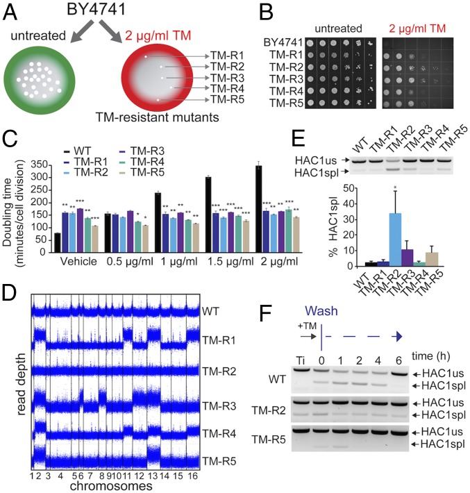
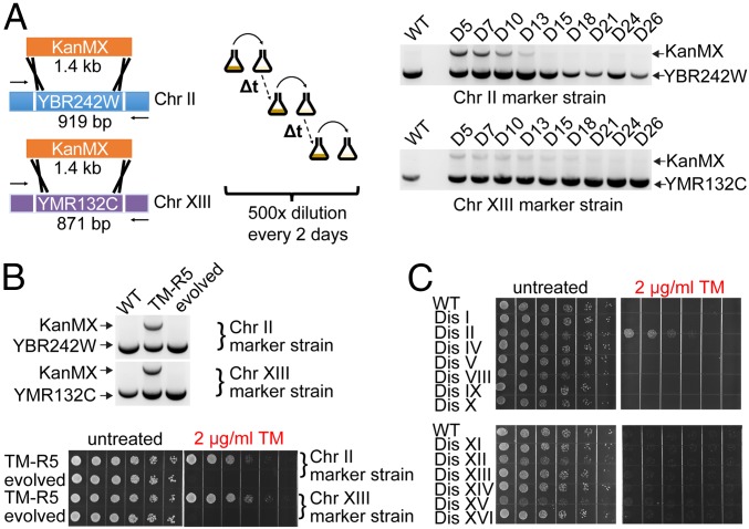
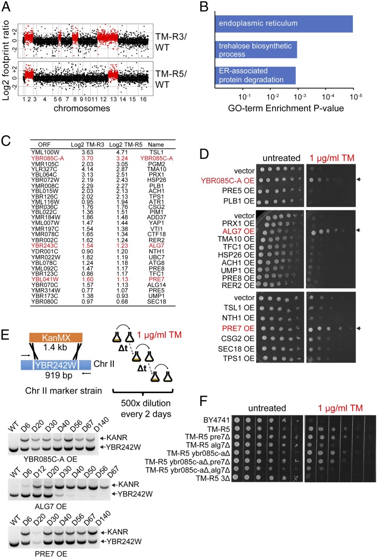
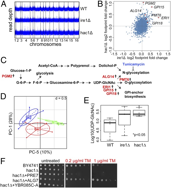
The yeast genome becomes unstable during stress, which often results in adaptive aneuploidy, allowing rapid activation of protective mechanisms that restore cellular homeostasis. In this study, we performed a genetic screen in Saccharomyces cerevisiae to identify genome adaptations that confer resistance to tunicamycin-induced endoplasmic reticulum (ER) stress. Whole-genome sequencing of tunicamycin-resistant mutants revealed that ER stress resistance correlated significantly with gains of chromosomes II and XIII. We found that chromosome duplications allow adaptation of yeast cells to ER stress independently of the unfolded protein response, and that the gain of an extra copy of chromosome II alone is sufficient to induce protection from tunicamycin. Moreover, the protective effect of disomic chromosomes can be recapitulated by overexpression of several genes located on chromosome II. Among these genes, overexpression of UDP-N-acetylglucosamine-1-P transferase (ALG7), a subunit of the 20S proteasome (PRE7), and YBR085C-A induced tunicamycin resistance in wild-type cells, whereas deletion of all three genes completely reversed the tunicamycin-resistance phenotype. Together, our data demonstrate that aneuploidy plays a critical role in adaptation to ER stress by increasing the copy number of ER stress protective genes. While aneuploidy itself leads to proteotoxic stress, the gene-specific effects of chromosome II aneuploidy counteract the negative effect resulting in improved protein folding.

Keywords: ER stress resistance; Saccharomyces cerevisiae; aneuploidy.


Conflict of interest statement

The authors declare no conflict of interest.

Figures

Fig. 1.
Fig. 1.
Genetic screen identifies gains of Chr II and XIII to be associated with resistance to ER stress. (A) Experimental design of the genetic screen used to isolate mutants that acquired resistance to TM-induced ER stress. (B) Representative images of wild-type (BY4741) cells and TM-resistant (TM-R) mutants grown in the presence of indicated concentrations of TM. Serial dilutions (10×) of logarithmically growing cells were spotted on control agar plates or plates with TM and incubated for 48 h at 30 °C. (C) Doubling time for TM-R strains in the presence of indicated concentrations of TM. Doubling time was calculated using the Yeast Outgrowth Data Analyzer software. Error bars represent SEM (n = 3). *P < 0.05, **P < 0.01, ***P < 0.001, Welch’s t test. (D) Duplication of chromosomes in TM-R strains was determined by whole-genome sequencing. Read depth was calculated in 100-bp windows. (E) Analysis of HAC1 mRNA splicing in TM-R mutants. Levels of spliced (spl) and unspliced (us) HAC1 mRNA were detected by RT-PCR. Error bars represent SEM (n = 3). *P < 0.05, Student’s t test. (F) Analysis of spliced HAC1 mRNA after TM washout in the TM-R2 and TM-R5 mutants. Logarithmically growing cells were treated with 1 μg/mL TM for 30 min; cells were washed and incubated in fresh media for indicated time. Levels of spliced (spl) and unspliced (us) HAC1 mRNA were detected by RT-PCR.
Fig. 2.
Fig. 2.
Chr II duplication is necessary and sufficient to confer ER stress resistance to cells. (A) The extra copies of Chr II or Chr XIII were labeled with the KanMX cassette. The laboratory evolution experiment was carried out by serial dilution in YPD medium. The loss of the extra Chr II or XIII was detected by PCR at the indicated days. (B) The loss of extra copies of the Chr II and XIII in the absence of TM reverted the TM resistance phenotype in the evolved TM-R5 populations. (C) Chr II duplication is sufficient to confer ER stress resistance to cells.
Fig. 3.
Fig. 3.
Overexpression of YBR085C-A, ALG7, and PRE7 in wild-type cells induces resistance to TM. (A) Translation levels of genes in TM-R3 and TM-R5 cells compared with a wild-type strain identified by Ribo-Seq. (B) Gene ontology enrichment analysis. Common genes up-regulated more than twofold in TM-R3 and TM-R5 mutants were analyzed using DAVID. (C) Common genes that were up-regulated more than 1.5-fold (0.6 in log2 scale) in both TM-R3 and TM-R5 mutants. Genes whose overexpression induces resistance to TM are shown in red. (D) Overexpression (OE) of YBR085C-A, ALG7, and PRE7 in wild-type cells induces resistance to TM. Resistance of strains to ER stress was determined using spot assays on SD agar plates containing 1 μg/mL TM. (E) ALG7 overexpression induces the loss of the extra Chr II in TM-R5 cells treated with TM. For the laboratory evolution experiment, the extra copy of Chr II in TM-R5 was labeled with the G418 resistance marker by replacing YBR242W with the KanMX cassette. TM-R5 marker strains overexpressing (OE) YBR085C-A, ALG7, or PRE7 were cultured in SD medium lacking leucine in the presence of 1 μg/mL TM. The laboratory evolution experiments were carried out by serial dilution with fresh medium every 2 d, and the loss of the extra copy of Chr II was detected by PCR at the indicated days. (F) Simultaneous deletion of extra copies of YBR085C-A, ALG7, and PRE7 (3Δ) prevents TM resistance in TM-R5 cells.
Fig. 4.
Fig. 4.
IRE1 and HAC1 deletion mutants activate enzymes involved in protein glycosylation and GPI anchor synthesis. (A) Genome coverage of the ire1Δ and hac1Δ mutants. Read depth was calculated in 100-bp windows. (B) Comparison of protein translation changes in the ire1Δ and hac1Δ mutants using Ribo-seq. Log2 footprint changes in the ire1Δ mutant compared with wild-type cells are plotted in x axis, and log2 footprint changes in the hac1Δ mutant are plotted in y axis. (C) Deletion of IRE1 and HAC1 genes activates protein glycosylation and GPI anchor synthesis. (D) Principal component analysis plot showing clustering of metabolites analyzed in the ire1Δ and hac1Δ mutants. (E) Analysis of UDP-GlcNAc levels. (F) Overexpression of ALG7 and PRE7, but not YBR085C-A, can rescue the growth of the hac1Δ mutant under ER stress.

References

    1. Parodi AJ. Role of N-oligosaccharide endoplasmic reticulum processing reactions in glycoprotein folding and degradation. Biochem J. 2000;348:1–13. - PMC - PubMed
    1. Walter P, Ron D. The unfolded protein response: From stress pathway to homeostatic regulation. Science. 2011;334:1081–1086. - PubMed
    1. Gardner BM, Pincus D, Gotthardt K, Gallagher CM, Walter P. Endoplasmic reticulum stress sensing in the unfolded protein response. Cold Spring Harb Perspect Biol. 2013;5:a013169. - PMC - PubMed
    1. Shamu CE, Walter P. Oligomerization and phosphorylation of the Ire1p kinase during intracellular signaling from the endoplasmic reticulum to the nucleus. EMBO J. 1996;15:3028–3039. - PMC - PubMed
    1. Aragón T, et al. Messenger RNA targeting to endoplasmic reticulum stress signalling sites. Nature. 2009;457:736–740. - PMC - PubMed

Publication types

MeSH terms

Substances

LinkOut - more resources