. 2018 Oct 29;92(22):e00825-18.
doi: 10.1128/JVI.00825-18. Print 2018 Nov 15.

PJA1 Coordinates with the SMC5/6 Complex To Restrict DNA Viruses and Episomal Genes in an Interferon-Independent Manner

Affiliations

PJA1 Coordinates with the SMC5/6 Complex To Restrict DNA Viruses and Episomal Genes in an Interferon-Independent Manner

Wei Xu et al. J Virol. .

Abstract

Viral and episomal DNAs, as signs of infections and dangers, induce a series of immune responses in the host, and cells must sense foreign DNAs to eliminate the invaders. The cell nucleus is not "immune privileged" and exerts intrinsic mechanisms to control nuclear-replicating DNA viruses. Thus, it is important to understand the action of viral DNA sensing in the cell nucleus. Here, we reveal a mechanism of restriction of DNA viruses and episomal plasmids mediated by PJA1, a RING-H2 E3 ubiquitin ligase. PJA1 restricts the DNA viruses hepatitis B virus (HBV) and herpes simplex virus 1 (HSV-1) but not the RNA viruses enterovirus 71 (EV71) and vesicular stomatitis virus (VSV). Similarly, PJA1 inhibits episomal plasmids but not chromosome-integrated reporters or endogenous genes. In addition, PJA1 has no effect on endogenous type I and II interferons (IFNs) and interferon-stimulated genes (ISGs), suggesting that PJA1 silences DNA viruses independent of the IFN pathways. Interestingly, PJA1 interacts with the SMC5/6 complex (a complex essential for chromosome maintenance and HBV restriction) to facilitate the binding of the complex to viral and episomal DNAs in the cell nucleus. Moreover, treatment with inhibitors of DNA topoisomerases (Tops) and knockdown of Tops release PJA1-mediated silencing of viral and extrachromosomal DNAs. Taken together, results of this work demonstrate that PJA1 interacts with SMC5/6 and facilitates the complex to bind and eliminate viral and episomal DNAs through DNA Tops and thus reveal a distinct mechanism underlying restriction of DNA viruses and foreign genes in the cell nucleus.IMPORTANCE DNA viruses, including hepatitis B virus and herpes simplex virus, induce a series of immune responses in the host and lead to human public health concerns worldwide. In addition to cytokines in the cytoplasm, restriction of viral DNA in the nucleus is an important approach of host immunity. However, the mechanism of foreign DNA recognition and restriction in the cell nucleus is largely unknown. This work demonstrates that an important cellular factor (PJA1) suppresses DNA viruses and transfected plasmids independent of type I and II interferon (IFN) pathways. Instead, PJA1 interacts with the chromosome maintenance complex (SMC5/6), facilitates the complex to recognize and bind viral and episomal DNAs, and recruits DNA topoisomerases to restrict the foreign molecules. These results reveal a distinct mechanism underlying the silencing of viral and episomal invaders in the cell nuclei and suggest that PJA1 acts as a potential agent to prevent infectious and inflammatory diseases.

Keywords: DNA topoisomerases; HBV; HSV-1; RING finger protein PJA1; SMC5/6 complex; episomal DNA; extrachromosomal DNA; hepatitis B virus; herpes simplex virus 1; host immune response.


Figures

FIG 1
FIG 1
PJA1 represses the transcription and replication of HBV and HSV-1. (A) Huh7 and HepG2 cells were plated in 24-well plates and transfected with 0.2 μg pHBV1.3 and 0.3 μg plasmids expressing 16 candidate proteins in triplicates for 48 h. (B and C) HepG2 cells (B) and Huh7 cells (C) were plated in 24-well plates and transfected with 0.2 μg pHBV1.3 and 0.3 μg pCAGGS-HA-PJA1 in triplicates for 48 h. HBeAg and HBsAg in the supernatants were assayed by an ELISA, and HBV pgRNA was measured by qPCR. (D) Diagrams of PJA1, PJA1B, PJA1ΔR, and PJA1BΔR proteins. (E) 293T cells were plated in 12-well plates and transfected with 1 μg pCAGGS-HA, pCAGGS-HA-PJA1, pCAGGS-HA-PJA1ΔR, pCAGGS-HA-PJA1B, and pCAGGS-HA-PJA1BΔR for 48 h. The expressed proteins were detected by Western blotting using anti-PJA1 antibody. (F) HepG2 cells were plated in 24-well plates overnight and transfected with 0.2 μg pHBV1.3 and 0.3 μg pCAGGS-HA, pCAGGS-HA-PJA1, and pCAGGS-HA-PJA1B for 48 h. HBeAg and HBsAg in the supernatants were assayed by an ELISA. (G) HepG2-NTCP cells were plated in 6-well plates, transfected with 2 μg pCAGGS-HA or pCAGGS-HA-PJA1B, and infected with HBV at 1,000 GE by inoculation with concentrated supernatants of HepaAD38 cells. HBeAg and HBsAg in the supernatants were assayed by an ELISA, and HBV pgRNA was measured by qPCR. OD450-630, optical density at 450 to 630 nm. (H) HepG2-NTCP cells were plated in 6-well plates, transfected with 2 μg pLKO.1-sh-NC or -sh-PJA1 for 24 h, and infected with HBV at 1,000 GE by inoculation with concentrated supernatants of HepaAD38 cells. sh-NC, nonspecific control shRNA. HBV pgRNA was measured by qPCR. Total DNA was extracted at 12 days postinfection (dpi), and DNA levels of HBV replication intermediates were measured by qPCR. (I and J) HepG2 cells (I) and Huh7 cells (J) were plated in 24-well plates and transfected with 0.3 μg pCAGGS-HA-PJA1B and 0.2 μg luciferase reporters containing the pre-S1, pre-S2, core, and X promoters of HBV for 48 h. Luciferase activities were determined, and results are expressed as fold induction relative to the control. (K) HepG2 cell lines stably expressing PJA1B were generated and infected with HSV-1 at an MOI of 0.1 for 8 h. HSV-1 US11 and ICP27 mRNA levels were determined by RT-qPCR. (L) HepG2-sh-NC and HepG2-sh-PJA1 cells were infected with HSV-1 at an MOI of 0.1 for 8 h. (Left) HSV-1 US11 and ICP27 mRNA levels were determined by RT-qPCR. (Right) HepG2 cell lines stably expressing pLKO.1-sh-NC or -sh-PJA1 were generated, and PJA1 mRNA levels in HepG2-sh-NC and HepG2-sh-PJA1 cells were detected. (M) Vero cells were plated in 6-well plates, transfected with 2 μg pCAGGS-HA or pCAGGS-HA-PJA1B for 24 h, and infected with HSV-1 at an MOI of 0.1. At 48 h postinfection, cell culture supernatants were collected, and the viral yields were determined by a plaque assay. Data are shown as means ± SD and correspond to results from a representative experiment out of three performed. **, P < 0.01; ***, P < 0.001.
FIG 2
FIG 2
PJA1 generally represses viral and episomal DNAs independent of IFN signaling pathways. (A and B) 293T cells (A) or HepG2 cells (B) were plated in 12-well plates and transfected with 1 μg pCAGGS-HA or pCAGGS-HA-PJA1B for 24 h. The mRNA levels of endogenous IFN-α, IFN-β, and IFN-γ genes were measured by RT-qPCR. (C to E) 293T cells were plated in 12-well plates; transfected with 1 μg pCAGGS-HA or pCAGGS-HA-PJA1B for 24 h; and treated with recombinant human IFN-α (rhIFN-α) (300 IU/ml), rhIFN-β (10 ng/ml), and rhIFN-γ (50 ng/ml) for 12 h. The mRNA levels of endogenous PKR (C), OAS1 (D), and MX1 (E) genes were measured by RT-qPCR. (F) HepG2 cells were plated in 12-well plates; transfected with 1 μg pCAGGS-HA or pCAGGS-HA-PJA1B for 24 h; and treated with rhIFN-α (300 IU/ml), rhIFN-β (10 ng/ml), and rhIFN-γ (50 ng/ml) for 12 h. The mRNA level of the endogenous PKR gene was measured by RT-qPCR. (G and H) 293T cells (G) or HepG2 cells (H) were treated with rhIFN-α (300 IU/ml), rhIFN-β (10 ng/ml), and rhIFN-γ (50 ng/ml) for 12 h. The PJA1 and GAPDH mRNA levels were measured by RT-qPCR. (I) 293T cells were plated in 24-well plates and cotransfected with 0.3 μg pCAGGS-HA or pCAGGS-HA-PJA1B and 0.2 μg reporters, pIFN-β-Luc, pISRE-Luc, pNF-κB-Luc, pTp53-Luc, and pCMV-Luc for 24 h. Luciferase activities were measured. (J) 293T cells were plated in 24-well plates and cotransfected with 0.3 μg pCAGGS-HA, pCAGGS-HA-PJA1, or pCAGGS-HA-PJA1B and 0.2 μg pCMV-Luc for 24 h. Luciferase activities were measured. (K to M) 293T cells were plated in 24-well plates and cotransfected with 0.2 μg pCMV-Luc and pCAGGS-HA-PJA1B at 0, 0.01, 0.05, and 0.1 μg for 24 h. (K) Luciferase activity was measured. (L) The Luc mRNA level was quantified by RT-qPCR. (M) pCMV-Luc DNA in the nuclear extract was quantified by qPCR. (N to P) 293T cells were plated in 24-well plates and cotransfected with 0.3 μg pCAGGS-HA, pCAGGS-HA-PJA1B, or pCAGGS-HA-PJA1BΔR and 0.2 μg reporters, pISRE-Luc (L), pCMV-Luc (M), and pTK-Renilla-Luc (N) for 24 h. Luciferase activities were measured. (Q and R) 293T cells were plated in 12-well plates and transfected with 0.5 μg pEGFP and 0.5 μg pCAGGS-HA, pCAGGS-HA-PJA1B, or pCAGGS-HA-PJA1BΔR for 24 h. (Q) GFP, PJA1, and β-actin were detected by Western blotting. WCL, whole-cell lysate. (R) Fluorescence intensity of EGFP was detected. Data are shown as means ± SD and correspond to results of a representative experiment out of three performed. Results are expressed as fold induction relative to the control. ns, not significant (P > 0.05); *, P ≤ 0.05; **, P < 0.01; ***, P < 0.001.
FIG 3
FIG 3
PJA1 has no effect on chromosome-integrated genes and RNA viruses. (A) 293T-Luc cells were generated, in which the CMV promoter driving Luc was randomly integrated into cell chromosomes by a lentiviral system. The cells were plated in 24-well plates and transfected with 0.5 μg pCAGGS, pCAGGS-HA-PJA1, or pCAGGS-HA-PJA1ΔR for 48 h. Luciferase activities were measured. Data are shown as means ± SD and correspond to results of a representative experiment out of three performed. ns, nonsignificant. (B) 293T-EGFP cells were generated, in which the CMV promoter driving EGFP was randomly integrated into cellular chromosomes by a lentiviral system. The cells were plated in 12-well plates and transfected with 1 μg pCAGGS, pCAGGS-HA-PJA1, or pCAGGS-HA-PJA1ΔR. GFP and β-actin protein were detected by Western blot analyses. (C) 293T cells were plated in 12-well plates and cotransfected with 0.5 μg pFlag-IRAK1 and 0.5 μg pCAGGS, pCAGGS-HA-PJA1, or pCAGGS-HA-PJA1ΔR for 48 h. Flag-IRAK1 and GAPDH protein levels were determined by Western blot analyses. (D) 293T cells were plated in 12-well plates and transfected with 1 μg pCAGGS, pCAGGS-HA-PJA1, or pCAGGS-HA-PJA1ΔR for 48 h. Endogenous IRAK1 and GAPDH were detected by Western blot analyses. (E) HepG2 cells were plated in 12-well plates and cotransfected with 0.5 μg pCAGGS-HA-IFNAR1 and 0.5 μg pcDNA3.1, pcDNA3.1-PJA1, or pcDNA3.1-PJA1ΔR for 48 h. HA-IFNAR1 and GAPDH were detected by Western blot analyses. (F) HepG2 cells were plated in 12-well plates and transfected with 1 μg pcDNA3.1, pcDNA3.1-PJA1, or pcDNA3.1-PJA1ΔR for 48 h. Endogenous IFNAR1 and GAPDH were detected by Western blot analyses. (G) RD cells were plated in 6-well plates, transfected with pCAGGS-HA-PJA1B at different concentrations (0, 0.5, 1, and 2 μg) for 48 h, and infected with EV71 at an MOI of 5 for 6 h. EV71 VP1, HA-PJA1B, and GAPDH were detected by Western blot analyses. (H) 293T cells were plated in 6-well plates, transfected with pCAGGS-HA-PJA1B at different concentrations (0, 0.5, 1, and 2 μg) for 24 h, and infected with VSV-GFP at an MOI of 1 for 12 h. GFP, PJA1B, and GAPDH were detected by Western blot analyses.
FIG 4
FIG 4
Knockout of PJA1 facilitates transcription of ectopic DNA. (A and B) To measure the effect of endogenous PJA1 on the regulation of episomal gene activation and DNA virus replication, the Cas9-mediated PJA1 knockout cell line 293T(PJA1-KO) and the control cell line with the pSpCas9-2A-Puro-MCS vector integrated were generated. Shown are bright-field images (A) and PJA1 protein levels (B) for 293T control cells and 293T(PJA1-KO) cells. (C) 293T control cells and 293T(PJA1-KO) cells were plated in a 96-well plate at a density of 1,000 cells/well in 100 μl of culture medium in triplicates. Ten microliters of a CCK-8 solution was added to each well of the plate, and the plate was incubated for 2 h in an incubator. The absorbance was measured at 450 nm. (D and E) 293T control cells and 293T(PJA1-KO) cells were plated in 24-well plates in triplicate and then transfected with 0.2 μg HBV Enh1-Luc (D) and 0.2 μg Tp53-Luc (E) for 24 h, and luciferase activity and relative luciferase mRNA levels were measured. (F) Tp53 protein levels in 293T control cells or 293T(PJA1-KO) cells were detected by Western blot analysis. (G) 293T control cells and 293T(PJA1-KO) cells were plated in 12-well plates and transfected with 0.2 μg pHBV-Enh1-Luc with or without 2 ng pCAGGS-HA-PJA1B for 24 h. Luciferase activities of HBV Enh1-Luc and protein levels of PJA1 and GAPDH were measured. (H) 293T control cells and 293T(PJA1-KO) cells were plated in 6-cm dishes and transfected with 5 μg pHBV-Enh1-Luc for 24 h. NSE4 binding to the episomal reporter or the GAPDH gene was monitored by ChIP assays using anti-NSE4 antibody. The amount of immunoprecipitated DNA was determined by RT-qPCR. (I and J) 293T control cells and 293T(PJA1-KO) cells were plated in 12-well plates overnight and then infected with HSV-1 at an MOI of 0.1 for the indicated times. HSV-1 US11 gene mRNA (I) and ICP27 gene mRNA (J) levels were determined by RT-qPCR. h.p.i, hours postinfection. Data are shown as means ± SD and correspond to results of a representative experiment out of three performed. ***, P < 0.001.
FIG 5
FIG 5
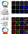
PJA1 interacts with the SMC5/6 complex in the nucleus. (A) Schematic of the SMC5/6 complex. (B) 293T cells were plated in 6-well plates and cotransfected with 1 μg pCAGGS-HA-PJA1ΔR and 1 μg pFlag, pFlag-SMC5, pFlag-SMC6, pFlag-NSE1, pFlag-NSE3, or pFlag-NSE4. Cells were lysed in RIPA lysis buffer. The immunoprecipitates and whole-cell lysates were analyzed by Western blotting with anti-HA or anti-Flag antibody. IB, immunoblot. (C) 293T cells were lysed in RIPA lysis buffer, and cell lysates were immunoprecipitated with anti-PJA1 antibody, anti-NSE4 antibody, or rabbit IgG. The immunoprecipitates and whole-cell lysates were analyzed by Western blotting with antibody to PJA1, NSE4, or GAPDH. (D) GST pulldown with cell lysates containing MBP-PJA1BΔR and GST, GST-NSE3, or GST-NSE4 expressed and purified from E. coli. After pulldown, precipitates were analyzed by Western blotting with anti-PJA1 or anti-GST antibody. (E and F) HepG2 cells were plated in confocal dishes and transfected with 1 μg pCAGGS-HA-PJA1, pFlag-SMC5, or pFlag-NSE4 (E) or cotransfected with 1 μg pCAGGS-HA-PJA1 and 1 μg pFlag-SMC5, pFlag-NSE3, pFlag-NSE4, or pFlag-DDB1 (F). Cells were immunostained with anti-HA and anti-Flag antibodies. Immunofluorescence analysis shows PJA1 (red), NSE3/4 (green), SMC5 (green), and DDB1 (green). The nucleus was stained by DAPI. (G) Schematic of the SMC5/6 complex in which NSE1 was replaced by PJA1.
FIG 6
FIG 6
PJA1 facilitates the binding of the SMC5/6 complex to viral and episomal DNAs. (A and B) 293T cells were plated in 6-cm dishes and cotransfected with 3 μg CMV-Luc and then with 2 μg pFlag-NSE1 (A) or 2 μg pcDNA3.1-PJA1B (B). NSE4 binding of the episomal reporter or GAPDH gene was monitored by ChIP assays using anti-NSE4 antibody. The level of immunoprecipitated DNA was determined by RT-qPCR. (C) 293T cells were plated in 6-well plates and cotransfected with 0.5 μg pCAGGS-HA-NSE4 and 0.5 μg pFlag-SMC5, pFlag-NSE1, or pFlag-NSE3 with or without 0.5 μg pcDNA3.1-PJA1ΔR. (D) 293T cells were plated in 6-well plates and cotransfected with 0.5 μg pCAGGS-HA-NSE4 and 0.5 μg pFlag-NSE1 and then with pcDNA3.1-PJA1BΔR at different concentrations (0, 0.5, and 1.5 μg). Cells were lysed in RIPA lysis buffer. The immunoprecipitates and whole-cell lysates were analyzed by Western blotting with anti-HA or anti-Flag antibody. (E and F) HepG2 cells stably expressing PJA1B were plated in 6-cm dishes and infected with HSV-1 at an MOI of 10 for 8 h. The binding of NSE4 to the VP16 gene promoter (E) or UL36 gene (F) of HSV-1 was monitored by a ChIP assay using beads only, rabbit IgG, or anti-NSE4 antibody. Data are shown as means ± SD and correspond to results of a representative experiment out of three performed. *, P < 0.05; **, P < 0.01; ***, P < 0.001.
FIG 7
FIG 7
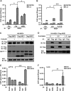
DNA topoisomerases are involved in PJA1-mediated restriction of viral and episomal DNAs. (A) 293T cells were plated in 12-well plates and transfected with 1 μg sh-NC, sh-SMC6, or sh-PJA1 for 48 h. SMC6, PJA1, NSE4, NSE1, and GAPDH proteins expressed in the transfected cells and in 293T control or 293T(PJA1-KO) cells were detected by Western blot analyses using the corresponding antibodies. WT, wild type. (B) 293T cells were plated in 24-well plates, cotransfected with 0.2 μg pCMV-Luc and 0.2 μg sh-SMC6 or sh-NC for 48 h, and then transfected with 0.2 μg pCAGGS or pCAGGS-HA-PJA1B for 48 h. Luciferase activities were measured. (C) Two stable cell lines were generated, in which sh-NC and sh-SMC6 were stably expressed in HepG2 cells. The stable cells were cotransfected with 0.1 μg pCMV-Luc and pCAGGS-HA-PJA1B at different concentrations (0, 0.02, 0.05, and 0.1 μg) for 48 h. Luciferase activities were measured. (D to G) 293T cells were plated in 24-well plates and cotransfected with 0.3 μg pCAGGS-PJA1B and 0.2 μg pHBV-Enh1-Luc for 24 h. The transfected cells were treated with different inhibitors (D); with topotecan or idarubicin at 5 μM for 10 h (E); or with topotecan (F) or idarubicin (G) at concentrations of 1, 5, and 10 μM for 10 h. Luciferase activity was measured, and results are expressed as primary data (left) and fold induction relative to the control (right). (H) 293T cells were plated in 24-well plates and transfected with 150 μM siR-NC, siR-Top1, siR-Top2a, or siR-Top2b for 48 h. The Top1 (left), Top2a (middle), Top2b (right), and GAPDH mRNA levels were determined by RT-qPCR, and results are expressed as fold induction relative to the control. (I) 293T cells were plated in 24-well plates; cotransfected with 0.1 μg pEnh1-Luc and 150 μM siR-NC, siR-Top1, siR-Top2a, or siR-Top2b for 24 h; and then transfected with 0.2 μg pCAGGS or pCAGGS-HA-PJA1B for 24 h. Luciferase activity was measured, and results are expressed as primary data (left) and fold induction relative to the control (right). Data are shown as means ± SD and correspond to results of a representative experiment out of three performed. ns, nonsignificant; *, P < 0.05; **, P < 0.01; ***, P < 0.001. (J and K) Diagrams of proposed models for PJA1 function. (J) Under normal conditions, the NSE1/NSE3/NSE4 subcomplex ensures that the SMC5/6 complex maintains the host chromosome. (K) In response to viral and episomal DNAs, PJA1 replaces NSE1 to form the PJA1/NSE3/NSE4 subcomplex, which converts the function of the SMC5/6 complex to the restriction of viral (V), extrachromosomal (E), and plasmid (P) DNA molecules.

Similar articles

Cited by

References

    1. El-Serag HB. 2012. Epidemiology of viral hepatitis and hepatocellular carcinoma. Gastroenterology 142:1264–1273. doi:10.1053/j.gastro.2011.12.061. - DOI - PMC - PubMed
    1. Ganem D, Varmus HE. 1987. The molecular biology of the hepatitis B viruses. Annu Rev Biochem 56:651–693. doi:10.1146/annurev.bi.56.070187.003251. - DOI - PubMed
    1. Roizman B, Knipe DM, Whitley RJ. 2013. Herpes simplex viruses, p 1823–1897. In Knipe DM, Howley PM, Cohen JI, Griffin DE, Lamb RA, Martin MA, Racaniello VR, Roizman B (ed), Fields virology, 6th ed Lippincott Williams & Wilkins, Philadelphia, PA.
    1. Schacker T. 2001. The role of HSV in the transmission and progression of HIV. Herpes 8:46–59. - PubMed
    1. Yoon WJ, Cho YD, Cho KH, Woo KM, Baek JH, Cho JY, Kim GS, Ryoo HM. 2008. The Boston-type craniosynostosis mutation MSX2 (P148H) results in enhanced susceptibility of MSX2 to ubiquitin-dependent degradation. J Biol Chem 283:32751–32761. doi:10.1074/jbc.M803183200. - DOI - PubMed

Publication types

MeSH terms

LinkOut - more resources