. 2018 Oct 29;92(22):e01259-18.
doi: 10.1128/JVI.01259-18. Print 2018 Nov 15.

Qualitative Differences in Capsidless L-Particles Released as a By-Product of Bovine Herpesvirus 1 and Herpes Simplex Virus 1 Infections

Affiliations

Qualitative Differences in Capsidless L-Particles Released as a By-Product of Bovine Herpesvirus 1 and Herpes Simplex Virus 1 Infections

Tiffany Russell et al. J Virol. .

Abstract

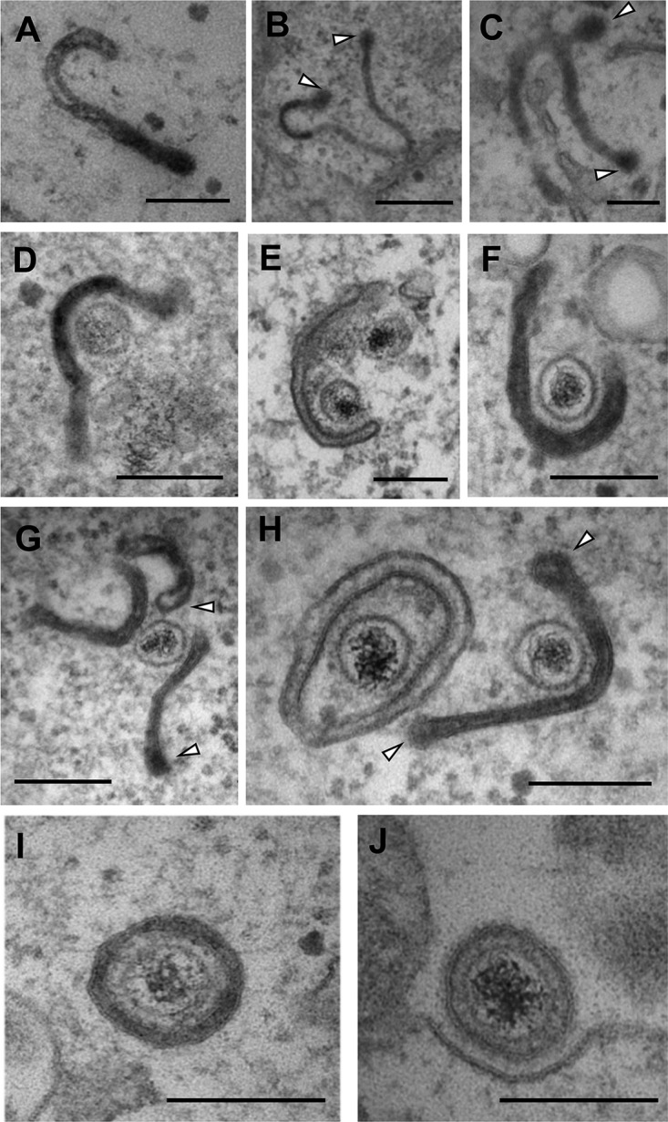
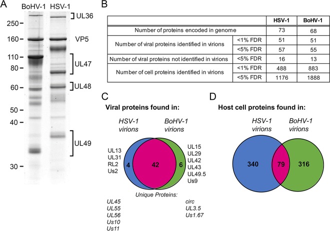
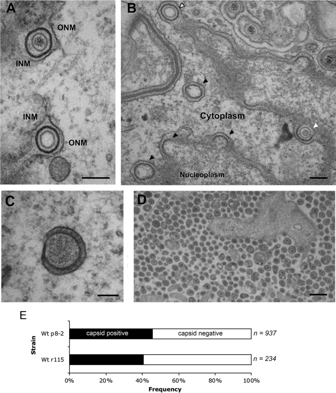
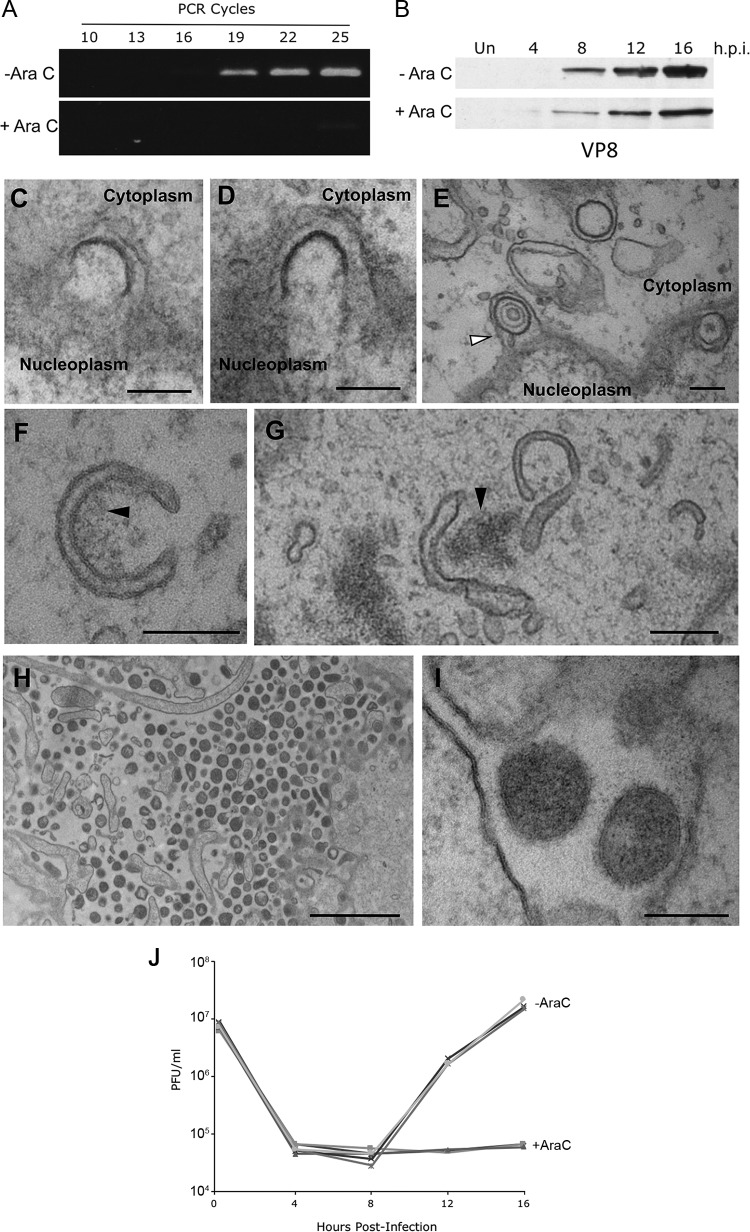
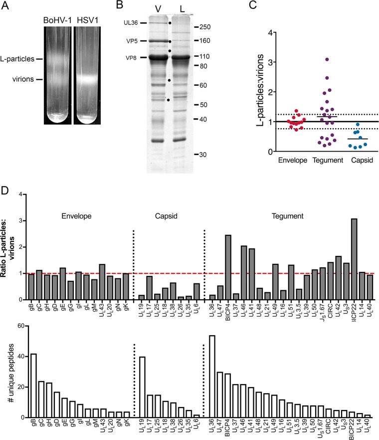
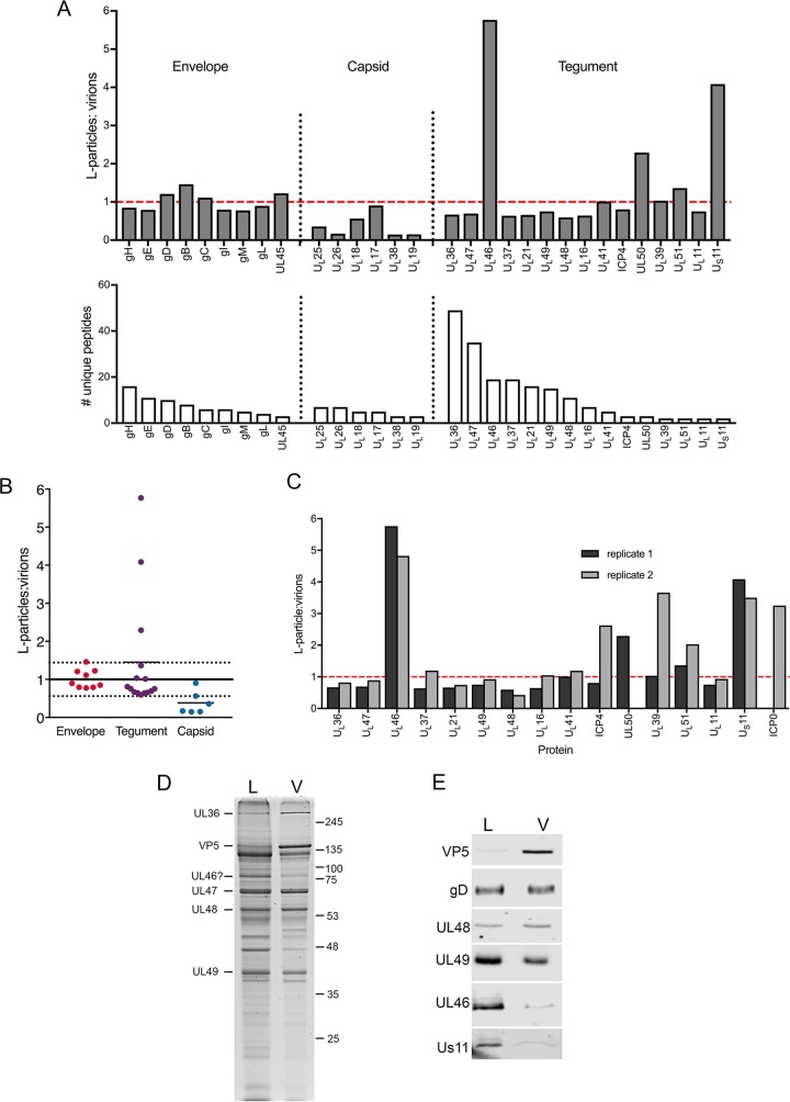
Despite differences in the pathogenesis and host range of alphaherpesviruses, many stages of their morphogenesis are thought to be conserved. Here, an ultrastructural study of bovine herpesvirus 1 (BoHV-1) envelopment revealed profiles similar to those previously found for herpes simplex virus 1 (HSV-1), with BoHV-1 capsids associating with endocytic tubules. Consistent with the similarity of their genomes and envelopment strategies, the proteomic compositions of BoHV-1 and HSV-1 virions were also comparable. However, BoHV-1 morphogenesis exhibited a diversity in envelopment events. First, heterogeneous primary envelopment profiles were readily detectable at the inner nuclear membrane of BoHV-1-infected cells. Second, the BoHV-1 progeny comprised not just full virions but also an abundance of capsidless, noninfectious light particles (L-particles) that were released from the infected cells in numbers similar to those of virions and in the absence of DNA replication. Proteomic analysis of BoHV-1 L-particles and the much less abundant HSV-1 L-particles revealed that they contained the same complement of envelope proteins as virions but showed variations in tegument content. In the case of HSV-1, the UL46 tegument protein was reproducibly found to be >6-fold enriched in HSV-1 L-particles. More strikingly, the tegument proteins UL36, UL37, UL21, and UL16 were depleted in BoHV-1 but not HSV-1 L-particles. We propose that these combined differences reflect the presence of truly segregated "inner" and "outer" teguments in BoHV-1, making it a critical system for studying the structure and process of tegumentation and envelopment.IMPORTANCE The alphaherpesvirus family includes viruses that infect humans and animals. Hence, not only do they have a significant impact on human health, but they also have a substantial economic impact on the farming industry. While the pathogenic manifestations of the individual viruses differ from host to host, their relative genetic compositions suggest similarity at the molecular level. This study provides a side-by-side comparison of the particle outputs from the major human pathogen HSV-1 and the veterinary pathogen BoHV-1. Ultrastructural and proteomic analyses have revealed that both viruses have broadly similar morphogenesis profiles and infectious virus compositions. However, the demonstration that BoHV-1 has the capacity to generate vast numbers of capsidless enveloped particles that differ from those produced by HSV-1 in composition implies a divergence in the cell biology of these viruses that impacts our general understanding of alphaherpesvirus morphogenesis.

Keywords: BoHV-1; HSV-1; L-particles; envelopment; morphogenesis; tegument.


Figures

FIG 1
FIG 1
BoHV-1 capsids are wrapped in tubular endocytic membranes. MDBK cells were infected at a multiplicity of infection (MOI) of 5 with BoHV-1, and HRP was added to the medium for 30 min at 12 h postinfection (hpi). Samples were fixed, processed, and imaged by transmission electron microscopy. White arrows in panels B, C, G, and H indicate the budded terminal domains of endocytic tubules. Bars = 500 nm (B) and 200 nm (A and C to J).
FIG 2
FIG 2
Purification and proteomic characterization of extracellular HSV-1 and BoHV-1 virions. Confluent monolayers of HaCaT cells and MDBK cells were infected with HSV-1 and BoHV-1, respectively; at full cytopathic effect, cells were harvested; and virions were isolated by separation on a 5 to 15% Ficoll gradient. (A) BoHV-1 and HSV-1 virions were separated by 10% SDS-PAGE and stained with Coomassie blue. Size markers are shown in kilodaltons. (B) Summary of the numbers of proteins identified in HSV-1 and BoHV-1 virions, at <5% and <1% FDRs. (C and D) Venn diagrams showing the overlap in virus proteins (C) and host cell proteins (D) identified in HSV-1 and BoHV-1 virions.
FIG 3
FIG 3
A variety of wrapping events occur in BoHV-1-infected cells. MDBK cells were infected at an MOI of 5 with BoHV-1 and fixed and processed for transmission electron microscopy at 12 hpi. (A and B) Representative sections of the nuclear envelope. In panel B, nuclear budding profiles that contain capsids resembling B capsids (white arrow) and A capsids (white arrow with black outline) or those occurring without a capsid (black arrows) are indicated. Bar = 200 nm. INM, inner nuclear membrane; ONM, outer nuclear membrane. (C) Fully wrapped capsidless L-particles were detected in the cytoplasm. Bar = 100 nm. (D) Multiple extracellular particles included full virions and capsidless L-particles. Bar = 500 nm. (E) Extracellular particles of MDBK cells infected with two strains of BoHV-1 were scored for the presence or absence of a capsid. Wt, wild type.
FIG 4
FIG 4
Inhibition of DNA replication with AraC does not arrest alternative wrapping events. (A and B) MDBK cells were infected at an MOI of 5 with BoHV-1 in the absence or presence of 100 μg/ml AraC. (A) The level of BoHV-1 genomic DNA was measured at 16 h by semiquantitative PCR using primers specific for UL47. (B) Synthesis of the late VP8 protein was measured by Western blotting of samples harvested at the indicated times. Un, uninfected. (C to I) MDBK cells were infected at an MOI of 5 with BoHV-1 in the presence of 100 μg/ml AraC. Samples were fixed and processed at 16 h, before processing for transmission electron microscopy. (C and D) Incomplete budding through the INM in the direction of the perinuclear space. (E) Several sealed vesicles are shown in the perinuclear space, with the arrow indicating a vesicle containing a B capsid. (F and G) Curved cytoplasmic tubules within the cytoplasm, frequently in association with electron-dense aggregates of material, indicated by the black arrows. (H and I) Extracellular space immediately adjacent to the plasma membrane, showing electron-dense particles, without a capsid cargo. Bars = 200 nm (C to G and I) and 2 μm (H). (J) MDBK cells were infected at an MOI of 5 with BoHV-1 in the absence (−AraC) or presence (+AraC) of 100 μg/ml AraC. Total virus was harvested at the indicated times and titrated on MDBK cells. Data from three biological replicates under each condition are shown.
FIG 5
FIG 5
Quantitative proteomic comparison of the L-particles and virions produced during BoHV-1 infection. (A) Confluent monolayers of MDBK cells were infected with BoHV-1 strain P8-2, and HaCaT cells were infected with HSV-1 strain Sc16, both at a multiplicity of 0.02. At full cytopathic effect, the extracellular medium was harvested, and released particles were isolated on 5% to 15% Ficoll gradients. Representative Ficoll gradients of BoHV-1 and HSV-1 particle preparations isolated at the same time are shown. (B) Equal amounts of the virion (V) and L-particle (L) bands of BoHV-1 were separated by 10% SDS-PAGE and stained with Coomassie blue. Black dots denote protein bands present in virions but not L-particles. Size markers are shown in kilodaltons. (C) The BoHV-1 virion and L-particle samples were subjected to TMT labeling and analyzed by mass spectrometry. Shown is a summary of the normalized ratios of virus proteins detected in BoHV-1 L-particles to virions, grouped according to their predicted location within the virion. The normalization line represents the average of all glycoprotein ratios, with 1 standard deviation on either side represented by dotted lines. (D) Ratio of individual virus proteins in BoHV-1 L-particles compared to virions (top), in order of the number of unique peptides detected (bottom). The dashed red line is the glycoprotein normalization line.
FIG 6
FIG 6
Quantitative proteomic comparison of the L-particles and virions produced in HSV-1 infection. (A) A Ficoll gradient for HSV-1 strain Sc16 similar to that shown in Fig. 5A was harvested for virions and L-particles as for BoHV-1 and analyzed by tandem mass tag labeling mass spectrometry as for BoHV-1. The results are presented as the ratio of the number of individual virus proteins in HSV-1 L-particles to virions (top), in order of the number of unique peptides detected (bottom). The dashed red line is the glycoprotein normalization line. (B) Summary of the normalized ratios of virus proteins detected in HSV-1 L-particles to virions, grouped according to their predicted location within the virion. The normalization line represents the average of all glycoprotein ratios, with 1 standard deviation on either side represented by dotted lines. (C) Comparative ratios of individual tegument proteins between two independent TMT labeling analyses. (D and E) Approximately equivalent amounts of Sc16 virions (V) and L-particles (L) were subjected to SDS-PAGE on a 10% polyacrylamide gel followed by Coomassie blue staining (D) or Western blotting (E) for the indicated virus proteins. Approximately equivalent numbers of particles were loaded by equating gD and UL48 by Western blotting. Molecular weight markers are shown in kilodaltons.

Similar articles

Cited by

References

    1. Looker KJ, Magaret AS, May MT, Turner KME, Vickerman P, Newman LM, Gottlieb SL. 2017. First estimates of the global and regional incidence of neonatal herpes infection. Lancet Glob Health 5:e300–e309. doi:10.1016/S2214-109X(16)30362-X. - DOI - PMC - PubMed
    1. Looker KJ, Magaret AS, May MT, Turner KME, Vickerman P, Gottlieb SL, Newman LM. 2015. Global and regional estimates of prevalent and incident herpes simplex virus type 1 infections in 2012. PLoS One 10:e0140765. doi:10.1371/journal.pone.0140765. - DOI - PMC - PubMed
    1. Statham JME, Randall LV, Archer SC. 2015. Reduction in daily milk yield associated with subclinical bovine herpesvirus 1 infection. Vet Rec 177:339. doi:10.1136/vr.103105. - DOI - PubMed
    1. Bennett R. 2003. The ‘direct costs’ of livestock disease: the development of a system of models for the analysis of 30 endemic livestock diseases in Great Britain. J Agric Econ 54:55–71. doi:10.1111/j.1477-9552.2003.tb00048.x. - DOI
    1. Muylkens B, Thiry J, Kirten P, Schynts F, Thiry E. 2007. Bovine herpesvirus 1 infection and infectious bovine rhinotracheitis. Vet Res 38:181–209. doi:10.1051/vetres:2006059. - DOI - PubMed

Publication types

LinkOut - more resources