. 2018 Nov 12;92(23):e01293-18.
doi: 10.1128/JVI.01293-18. Print 2018 Dec 1.

A Proteomic Atlas of the African Swine Fever Virus Particle

Affiliations

A Proteomic Atlas of the African Swine Fever Virus Particle

Alí Alejo et al. J Virol. .

Abstract

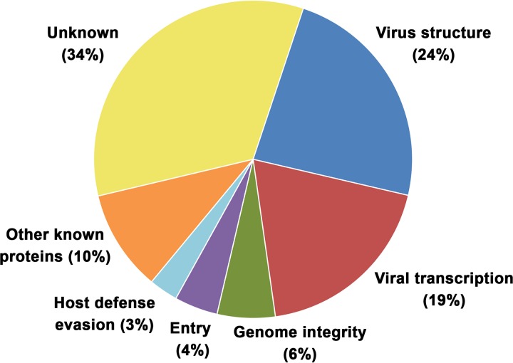
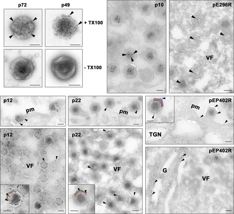
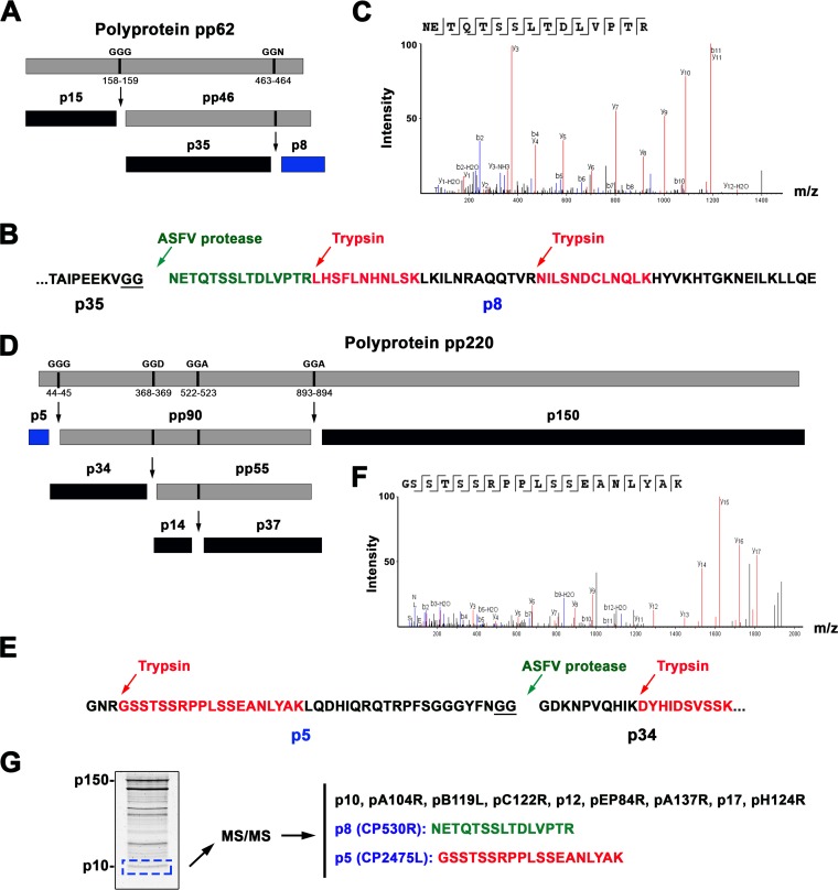
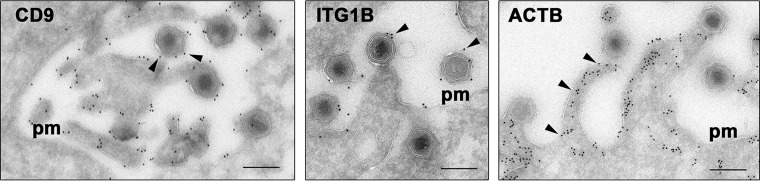
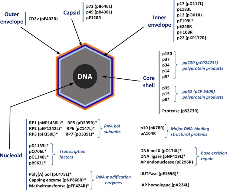
African swine fever virus (ASFV) is a large and complex DNA virus that causes a highly lethal swine disease for which there is no vaccine available. The ASFV particle, with an icosahedral multilayered structure, contains multiple polypeptides whose identity is largely unknown. Here, we analyzed by mass spectroscopy the protein composition of highly purified extracellular ASFV particles and performed immunoelectron microscopy to localize several of the detected proteins. The proteomic analysis identified 68 viral proteins, which account for 39% of the genome coding capacity. The ASFV proteome includes essentially all the previously described virion proteins and, interestingly, 44 newly identified virus-packaged polypeptides, half of which have an unknown function. A great proportion of the virion proteins are committed to the virus architecture, including two newly identified structural proteins, p5 and p8, which are derived from the core polyproteins pp220 and pp62, respectively. In addition, the virion contains a full complement of enzymes and factors involved in viral transcription, various enzymes implicated in DNA repair and protein modification, and some proteins concerned with virus entry and host defense evasion. Finally, 21 host proteins, many of them localized at the cell surface and related to the cortical actin cytoskeleton, were reproducibly detected in the ASFV particle. Immunoelectron microscopy strongly supports the suggestion that these host membrane-associated proteins are recruited during virus budding at actin-dependent membrane protrusions. Altogether, the results of this study provide a comprehensive model of the ASFV architecture that integrates both compositional and structural information.IMPORTANCE African swine fever virus causes a highly contagious and lethal disease of swine that currently affects many countries of sub-Saharan Africa, the Caucasus, the Russian Federation, and Eastern Europe and has very recently spread to China. Despite extensive research, effective vaccines or antiviral strategies are still lacking, and many basic questions on the molecular mechanisms underlying the infective cycle remain. One such gap regards the composition and structure of the infectious virus particle. In the study described in this report, we identified the set of viral and host proteins that compose the virion and determined or inferred the localization of many of them. This information significantly increases our understanding of the biological and structural features of an infectious African swine fever virus particle and will help direct future research efforts.

Keywords: African swine fever virus; NCLDV; giant virus; immunoelectron microscopy; mass spectrometry; proteome; proteomic analysis; virus composition; virus structure.


Figures

FIG 1
FIG 1
Quality control of purified ASFV particles. (A) Percoll-purified ASFV particles were analyzed by electron microscopy after negative staining. Note that ASFV virions look intact and essentially free of contaminant structures. Bar, 100 nm. (B) The virion proteins were analyzed by SDS-PAGE and stained with colloidal Coomassie blue. Some of the most prominent protein bands are indicated. (C) Western immunoblotting of extracts of mock-infected cells (lanes M) and ASFV-infected cells (lanes I) as well as purified ASFV (lanes V) with antibodies against the structural protein p150, derived from polyprotein pp220, and p35, derived from polyprotein pp62. Note the absence of the polyprotein precursors in the virus fraction, which are present in the infected cell extract.
FIG 2
FIG 2
Functional classification of ASFV virion proteins. The viral proteins identified in the ASFV proteome are assigned to the following functional categories: virus structure and morphogenesis (p72, p150, p37, p34, p14, p5, p35, p15, p8, pS273R, p17, pE183L, p49, pE120R, p10, and pA104R), viral transcription and RNA modification (pB962L, pD1133L, pNP1450L, pEP1242L, pG1340L, pQ706L, pNP868R, pC475L, pH359L, pD205R, pD339L, pEP424R, and pC147L), maintenance of genome integrity (pO174L, pNP419L, pE296R, and pE165R), virus entry (p12, pE248R, and pE199L), evasion from host defense (pEP402R and pA224R), other known proteins (pR298L, pB119L, p22, p32, p11.5, pEP152R, and pH339R), and unknown proteins (pM1249L, pCP123L, pC129R, pC717R, pI177L, pK145R, pK421R, pE146L, pF317L, pH240R, pCP312R, pE423R, pE184L, pC257L, pH171R, pB117L, pB169L, pEP84R, pI73R, pC122R, pQP383R, pM448R, and pH124R).
FIG 3
FIG 3
Immunoelectron microscopy of viral proteins in the virus particle. To localize protein p49, ASFV particles were treated or not with 0.1% Triton X-100 (TX100) and incubated with a control antibody against the major capsid protein p72 or an antibody against p49, followed by protein A-gold (5 nm). Note that both anti-p72 and anti-p49 antibodies stained the capsid on detergent-treated particles. To localize proteins p10, pE296R, p12, p22, and pEP402R, cryosections of ASFV-infected cells were incubated with specific antibodies followed by protein A-gold (10 nm). Note that both anti-p10 and anti-pE296R labeled the nucleoid region, whereas the anti-p12 and anti-p22 antibodies labeled the inner viral envelope of virus particles at the virus factories (VF) and the plasma membrane (pm). The anti-pEP402R serum mainly stained the Golgi stack (G) as well as presumed trans-Golgi network membranes. A minor fraction of pEP402R was also found on the plasma membrane and on the outer viral envelope of budding ASFV particles. (Insets) Details of labeling of intracellular (p12 and p22) and extracellular (pEP402R) particles. The arrowheads indicate, for each case, examples of the described distribution patterns. To facilitate the interpretation, the inner membrane (red), capsid (green), and outer membrane (purple) are depicted in color. Bars, 100 nm.
FIG 4
FIG 4
Two novel structural ASFV proteins, p8 and p5, are derived from the core polyproteins pp62 and pp220, respectively. (A) Schematic representation of polyprotein pp62 processing at GGX cleavage sites. The polyprotein pp62, the intermediate precursor pp46, and the mature products p15, p35, and p8 (in blue) are indicated. (B) Detection of p8 in the ASFV proteome. Protein p8 was identified by two tryptic peptides (highlighted in red) and one peptide (green) matching the N-terminal p8 sequence, which is generated by cleavage by the ASFV proteinase at positions 463 and 464 of the GGR site. (C) MS/MS spectrum of the N-terminal nontryptic p8 peptide NETQTSSLTDLVPTR. (D) Schematic representation of polyprotein pp220 processing at GGX cleavage sites. The polyprotein pp220, the intermediate precursors pp90 and pp55, and the mature products p5 (in blue), p34, p14, p150 are indicated. (E) Detection of protein p5 through one tryptic peptide (in red) covering 43% of its amino acid sequence. (F) MS/MS spectrum of the tryptic peptide GSSTSSRPPLSSEANLYAK. (G) Proteomic analysis of the low-molecular-weight virion proteins. ASFV proteins were separated by SDS-PAGE, and the low-molecular-weight region was excised and in-gel digested with trypsin. MS/MS analysis identified all the small viral proteins, including p8 and p5.
FIG 5
FIG 5
Immunoelectron microscopy of host proteins recruited to the virus particle. Thawed cryosections of ASFV-infected cells were stained with mouse MAbs against the tetraspanin CD9, integrin beta 1 (ITGB1), and beta-actin (ACTB), followed by rabbit anti-mouse immunoglobulins and protein A-gold (10 nm). Note that anti-CD9 and integrin beta 1 antibodies decorated the plasma membrane (pm) and the outer envelope of the virus particles released by budding. Note also that antiactin antibodies stained the membrane protrusions induced by the budding ASFV particles. The arrowheads indicate, for each case, examples of the described distribution patterns. Bars, 200 nm.
FIG 6
FIG 6
ASFV atlas. The subviral localization of 40 viral proteins among the five structural domains of the ASFV particle is shown. The distribution of proteins marked with an asterisk was inferred from the predicted or known role, while that of the remaining proteins was determined by immunoelectron microscopy. pol, polymerase.

Similar articles

Cited by

References

    1. Sanchez-Cordon PJ, Montoya M, Reis AL, Dixon LK. 2018. African swine fever: A re-emerging viral disease threatening the global pig industry. Vet J 233:41–48. doi:10.1016/j.tvjl.2017.12.025. - DOI - PMC - PubMed
    1. Zhou X, Li N, Luo Y, Liu Y, Miao F, Chen T, Zhang S, Cao P, Li X, Tian K, Qiu H, Hu R. 13 August 2018. Emergence of African swine fever in China, 2018. Transbound Emerg Dis. doi:10.1111/tbed.12989. - DOI - PubMed
    1. Iyer LM, Aravind L, Koonin EV. 2001. Common origin of four diverse families of large eukaryotic DNA viruses. J Virol 75:11720–11734. doi:10.1128/JVI.75.23.11720-11734.2001. - DOI - PMC - PubMed
    1. Iyer LM, Balaji S, Koonin EV, Aravind L. 2006. Evolutionary genomics of nucleo-cytoplasmic large DNA viruses. Virus Res 117:156–184. doi:10.1016/j.virusres.2006.01.009. - DOI - PubMed
    1. Yanez RJ, Rodriguez JM, Nogal ML, Yuste L, Enriquez C, Rodriguez JF, Vinuela E. 1995. Analysis of the complete nucleotide sequence of African swine fever virus. Virology 208:249–278. doi:10.1006/viro.1995.1149. - DOI - PubMed

Publication types

LinkOut - more resources