Review
. 2018 Sep;8(9):180112.
doi: 10.1098/rsob.180112.

Structural insights into the functional diversity of the CDK-cyclin family

Affiliations
Review

Structural insights into the functional diversity of the CDK-cyclin family

Daniel J Wood et al. Open Biol. 2018 Sep.

Abstract

Since their characterization as conserved modules that regulate progression through the eukaryotic cell cycle, cyclin-dependent protein kinases (CDKs) in higher eukaryotic cells are now also emerging as significant regulators of transcription, metabolism and cell differentiation. The cyclins, though originally characterized as CDK partners, also have CDK-independent roles that include the regulation of DNA damage repair and transcriptional programmes that direct cell differentiation, apoptosis and metabolic flux. This review compares the structures of the members of the CDK and cyclin families determined by X-ray crystallography, and considers what mechanistic insights they provide to guide functional studies and distinguish CDK- and cyclin-specific activities. Aberrant CDK activity is a hallmark of a number of diseases, and structural studies can provide important insights to identify novel routes to therapy.

Keywords: cell cycle; cyclin; kinase; transcription.


Conflict of interest statement

The authors declare that there are no competing interests associated with the manuscript. Some work in the authors' laboratory is funded by Astex Pharmaceuticals.

Figures
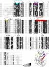
Figure 1.
Figure 1.
Sequence alignment of the human CDK family. Greyscale shading denotes the extent of sequence conservation calculated from UniProt sequences using Clustal Omega [33] and exported into ExPASy BoxShade. Structural features described in the text are named and highlighted in colour above the alignment and located on the fold of CDK1 (extracted from the CDK1–Cks1 complex, PDB code: 4YC6). UniProt codes used: CDK1 (P06493), CDK2 (P24941), CDK3 (Q00526), CDK4 (P11802), CDK5 (Q00535), CDK6 (Q00534), CDK7 (P50613), CDK8 (P49336), CDK9 (P50750), CDK10 (Q15131), CDK11A (Q9UQ88), CDK11B (P21127), CDK12 (Q9NYV4), CDK13 (Q14004), CDK14 (O94921), CDK15 (Q96Q40), CDK16 (Q00536), CDK17 (Q00537), CDK18 (Q07002), CDK19 (Q9BWU1), CDK20 (Q8IZL9). CDK11A and CDK11B result from a gene duplication and are almost identical (97.5%).
Figure 2.
Figure 2.
The monomeric CDK fold. (a) Structure of monomeric CDK2. The CDK kinase fold, as first exemplified by monomeric CDK2 ([39], PDB 1HCK), is composed of a smaller N-terminal lobe that is predominantly a twisted anti-parallel β-sheet linked via a flexible hinge sequence to a larger C-terminal lobe dominated in structure by α-helices (light blue ribbon). Structural features are highlighted: glycine-rich loop (sequence GXGXXG, cyan), αC-helix (residues 45–55, purple), hinge (residues 80–84, yellow), activation loop (residues 145–172, red). The location of T160 is marked. (b) The monomeric CDK fold is conserved as shown by an overlay of CDK1 (extracted from the structure of CDK1–Cks2), CDK2, CDK6, CDK7 and CDK16 structures. The other CDK folds are superposed on CDK2: CDK1 (PDB 4YC6, light grey); CDK6 (PDB 5L2S, cyan); CDK7 (PDB 1UA2, magenta) and CDK16 (PDB 5G6 V, light green). Mobility is indicated by the quality of the experimental electron density maps, so that the derived structures can be traced with varying degrees of confidence. (c) The various conformations the activation and glycine-rich loops can adopt are highlighted by this structural comparison. Structures reported for these loops may represent more populous low energy conformations compatible with a particular crystal lattice. This model is supported by studies of monomeric CDK2 phosphorylated on the conserved threonine residue within the activation loop (T160 in CDK2), which exhibits approximately 0.3% of the fully active CDK2–cyclin A complex ([40], PDB 1QMZ). The majority of the CDK2 probably corresponds to inactive conformations, but a small fraction is in an active conformation and generates the basal activity observed.
Figure 3.
Figure 3.
CDK activation by cyclin binding. (a) Overlay of monomeric CDK2 and T160-phosphorylated CDK2–cyclin A. Cyclin A composed of two tandem cyclin box folds (CBFs [49], PDB 1VIN) acts as a scaffold to which the malleable unphosphorylated CDK responds to generate a binary complex that exhibits basal activity ([47], PDB 1JST). The CDK αC-helix is rotated and relocated into the active site by engagement with the N-CBF of the cyclin subunit. At the start of the activation loop, αL12 is melted and the conserved DFG motif adopts an active ‘DFG-in’ conformation in which the aspartate side chain coordinates a magnesium ion to productively orientate the ATP phosphate groups for catalysis. The activation loop is extended and pulled away from the active site to form a platform that will ultimately recognize the protein substrate around the site of phospho-transfer ([50], PDB 1QMZ). Cyclin binding also refines the relative positions of the CDK2 N- and C-terminal lobes, so that residues within the hinge and lining the active site orientate the ATP adenine and ribose rings and phosphate groups for catalysis. Overall, the CDK2–cyclin A interface is extensive (2839 Å2, [51]) extending between both lobes of the CDK and the two cyclin CBFs, further strengthened by engagement of the cyclin N-terminal helix preceding the N-CBF with the CDK C-terminal lobe. The phospho-threonine within the activation loop (T160 in CDK2) acts as a structural hub liganded by conserved, positively charged residues located within the C-helix (R50), at the start of the activation loop (R150) and adjacent to the catalytic aspartate residue (R126). In the absence of T160 phosphorylation, a conserved C-terminal glutamate residue (E162 in CDK2) satisfies the positively charged side chains of the phospho-threonine-binding pocket, and the side chain hydroxyl of T160 is solvent accessible within the context of a relatively well-ordered activation loop ([47], PDB 1JST). The inactive conformation of CDK2 is shown as a translucent ribbon. The N-CBF and C-CBF are also shown. (b) CDK1–cyclin B (PDB 4YC3; CDK1 grey, cyclin B translucent cyan surface). Inactive (cyclin-unassociated) CDK1 conformation shown as a translucent ribbon. (c) CDK2–Spy1 is shown in a similar pose (PDB 5UQ2; CDK2 blue, Spy1 translucent pink surface). (d) Comparison of unphosphorylated CDK2–cyclin A (PDB 1FIN; activation loop, red), T160-phosphorylated CDK2–cyclin A with peptide present (PDB 2CCI; peptide, yellow activation loop, deep red) and CDK2–Spy1 (PDB 5UQ2; activation loop in brown) activation loop conformations. The positions of residues (P−3 to P+3) within the CDC6 peptide substrate (sequence HHASPRK) with respect to the serine residue at the site of phospho-transfer (P position) are indicated.
Figure 4.
Figure 4.
CDK–cyclin complexes. A comparison of the CDK–cyclin complexes, for which structures are available, highlights the differences in the CDK response to cyclin association. (a) CDK6–viral cyclin (PDB 1JOW, CDK6, cyan with activation loop (residues 163–189) shown in red; viral cyclin, grey). (b) CDK4–cyclin D1 (PDB 2W96, CDK4, orange; cyclin D1, light purple, RXL-binding site shown as a red translucent surface (residues 54–61) and partially resolved LXCXE motif shown in cyan (residues 6–9)). (c) CDK4–cyclin D3 (PDB 3G33, CDK4, orange; cyclin D3, purple, RXL-binding site shown as a red translucent surface (residues 56–61). (d) CDK5–p25 (PDB 1H4 L, CDK5, light blue with activation loop (residues 144–171) shown in red; p25, gold). (e) CDK8–cyclin C (CDK8, green with C-terminal residues 343–353 in orange; cyclin C, purple). (f) CDK9–cyclin T1 (PDB 3BLH, CDK9 lilac with C-terminal residues 317–325 in orange; cyclin T, pale yellow). (g) CDK12–cyclin K (PDB 4UN0, CDK12, light grey, C-terminal rail residues 1025–1036 in orange; cyclin K, green). (h) CDK13–cyclin K (PDB 5EFQ, CDK13, gold, C-terminal tail residues 1011–1025 in orange; cyclin K, green). The activation segment sequences are shown in red where resolved in the structures.
Figure 5.
Figure 5.
Shaping of the catalytic cleft by the C-terminal tail in the transcriptional CDKs. In each case, the binding of an ATP-competitive inhibitor (ball and stick model) within the ATP-binding pocket helps to order the C-terminal sequence. (a) CDK8–cyclin C–CCT251545 (PDB entry 5BNJ); (b) CDK9–cyclin T1–DRB (PDB 3MY1) overlaid with the full-length CKD9 structure (PDB 4RC8); (c) CDK12–cyclin K–THZ531 (PDB 5ACB). In this structure, the inhibitor THZ531 forms an irreversible bond with C1039 located within the CDK12 C-terminal extension. The CDK8, 9 and 12 folds are coloured green, lilac and grey, respectively, and the CDK C-terminal tails are coloured orange. The hinge region between the N- and C-terminal kinase lobes and the αC-helix is identified to provide the context.
Figure 6.
Figure 6.
CDK–cyclin interaction partners. A number of CDK–cyclin partners and interaction sites have also been solved structurally. (a) CDK2–cyclin A p27KIP1 (PDB 1JST, CDK2–cyclin A, coloured as previous, p27KIP1 is coloured green and the hydrophobic patch of the RXL site is highlighted in orange with p27KIP1 side chains R30, N31, L32, F33 highlighted). (b) CDK6–p19INK4d (PDB 1BLX, CDK6, cyan; p19INK4D, orange). (c) CDK1–Cks1 (PDB 4YC6, CDK1, grey; CKS1, blue with phospho-threonine (pT)-interacting residues shown in purple; the peptide from 2CCI (yellow) has been superposed onto 4YC6). (d) CDK2-KAP (PDB 1FQ1, CDK2, blue with red activation loop; KAP, green). (e) cyclin E–Fbw7 (PDB 2OVQ, Fbw7, orange; cyclin E peptide, green). (f) cyclin D1–FBXO31 (PDB 5VZU, FBX031, crimson; cyclin D1 peptide, pink).
Figure 7.
Figure 7.
The RXL substrate recruitment site is unavailable in transcriptional cyclins. Differences between the cell cycle and transcriptional cyclin families within the N-terminal cyclin box fold (N-CBF) reveal different availabilities of the RXL site. (a) The cyclin A RXL site with CDC6 (yellow, PDB 2CCI) and RB-associated protein (pink, PDB 1H25) peptides bound is unavailable in transcriptional cyclins (b) cyclin T (PDB 3BLH, orange), (c) cyclin K (PDB 4UN0, green) and (d) cyclin H (PDB 1KXU, blue) due to extended α4 helices and protruding α4–α5 loops.
Figure 8.
Figure 8.
CDK9–cyclin T binds Tat and AFF4. (a) The HIV Tat protein binds to the C-terminal cyclin box fold (C-CBF) of cyclin T (PDB 3MI9, CDK9–cyclin T-coloured as previous; Tat, blue). Tat contains an acidic-/proline-rich region and a cysteine-rich region for the coordination of Zn, with the second site completed by cyclin T C261. (b) CDK9–cyclin T-Tat also binds AFF4 at the C-CBF (PDB 4OGR, AFF4, red).
Figure 9.
Figure 9.
Cyclin D sequence conservation. (a) Sequence conservation between the cyclin D isoforms has been mapped onto the structure of CDK4–cyclin D1 (PDB 2W96) and is represented by blue-scale colouring. (b) Alignment of cyclin D1/2/3 conducted in Clustal Omega and output into ExPASy BoxShade. Secondary structure elements for cyclin D1 are shown above the sequence. CDK4 is coloured orange. The UniProt codes used for sequence alignment are: cyclin D1 (P24385), cyclin D2 (P30279) and cyclin D3 (P30281).
Figure 10.
Figure 10.
Comparison of cyclin T, cyclin D and cyclin A structures in the vicinity of the AFF4-binding site. (a) CDK2–cyclin A showing the C-CBF with the C-terminal cyclin A tail (orange) accommodated (PDB 1FIN, CDK2–cyclin A coloured as previous). The same site is presented for (b) cyclin D1 (PDB 2W96, coloured as previous) and (c) cyclin T (PDB 4OGR, coloured as previous), which is known to accommodate the binding of both AFF4 and Tat.
Figure 11.
Figure 11.
Point mutations in CDK4/6–p16INK4A. CDK4/6 and p16INK4A (represented here by CDK6, PDB 1BI7, CDK6, cyan; p16INK4A, gold) contain a number of residues that are frequently mutated in cancer. Several commonly described mutations occur on the CDK–p16INK4A interface, disrupting the complex and leading to kinase dysregulation. Other p16INK4A residues such as H98 and P48 are located further from the CDK-binding interface. CDK6 mutations are selected from those described in cancer genome repositories (see the text for further details).
Figure 12.
Figure 12.
The cryo-EM structure of the CDK4–Cdc37–Hsp90 complex. Both copies of Hsp90 are coloured blue; Cdc37 is drawn in red and CDK4 in orange. Structure is drawn from PDB 5FWK.

References

    1. Morgan DO. 2007. The cell cycle principles of control (primers in biology). London, UK: New Science Press.
    1. Malumbres M, Harlow E, Hunt T, Hunter T, Lahti JM, Manning G, Morgan DO, Tsai LH, Wolgemuth DJ. 2009. Cyclin-dependent kinases: a family portrait. Nat. Cell Biol. 11, 1275–1276. (10.1038/ncb1109-1275) - DOI - PMC - PubMed
    1. Malumbres M. 2014. Cyclin-dependent kinases. Genome Biol. 15, 122 (10.1186/gb4184) - DOI - PMC - PubMed
    1. Jeronimo C, Collin P, Robert F. 2016. The RNA polymerase II CTD: the increasing complexity of a low-complexity protein domain. J. Mol. Biol. 428, 2607–2622. (10.1016/j.jmb.2016.02.006) - DOI - PubMed
    1. Fisher RP, Morgan DO. 1994. A novel cyclin associates with MO15/CDK7 to form the CDK-activating kinase. Cell 78, 713–724. (10.1016/0092-8674(94)90535-5) - DOI - PubMed