. 2018 Sep 5;8(1):13263.
doi: 10.1038/s41598-018-31376-8.

Peptidyl arginine deiminase 2 (Padi2) is expressed in Sertoli cells in a specific manner and regulated by SOX9 during testicular development

Affiliations

Peptidyl arginine deiminase 2 (Padi2) is expressed in Sertoli cells in a specific manner and regulated by SOX9 during testicular development

Atsumi Tsuji-Hosokawa et al. Sci Rep. .

Abstract

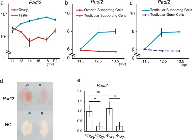
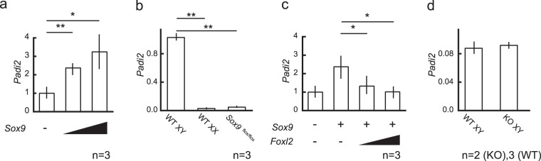
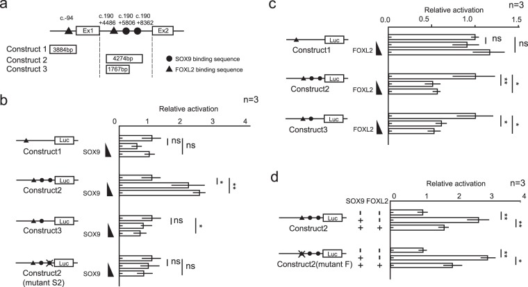
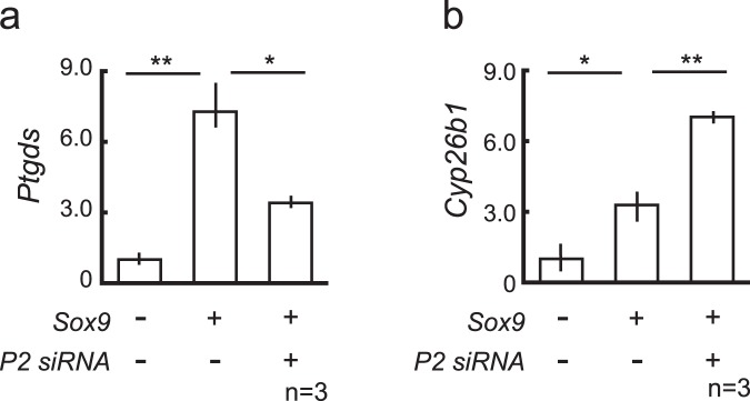
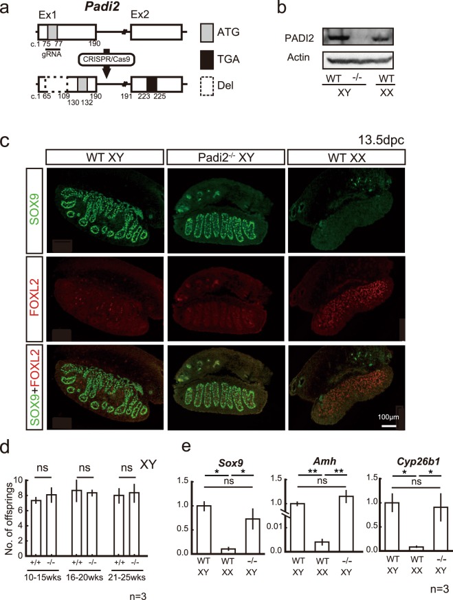
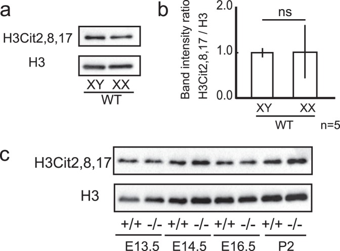
Peptidyl arginine deiminases (PADIs) are enzymes that change the charge of proteins through citrullination. We recently found Padi2 was expressed exclusively in fetal Sertoli cells. In this study, we analyzed the transcriptional regulation of Padi2 and the role of PADI2 in testicular development. We showed SOX9 positively regulated Padi2 transcription and FOXL2 antagonized it in TM3 cells, a model of Sertoli cells. The responsive region to SOX9 and FOXL2 was identified within the Padi2 sequence by reporter assay. In fetal testes from Sox9 knockout (AMH-Cre:Sox9flox/flox) mice, Padi2 expression was greatly reduced, indicating SOX9 regulates Padi2 in vivo. In vitro analysis using siRNA suggested PADI2 modified transcriptional regulation by SOX9. However, Padi2-/- XY mice were fertile and showed no apparent reproductive anomalies. Although, PADI2 is known as an epigenetic transcriptional regulator through H3 citrullination, no significant difference in H3 citrullination between wildtype and Padi2-/- XY gonads was observed. These results suggest Padi2 is a novel gene involved in testis development that is specifically expressed in Sertoli cells through the regulation by SOX9 and FOXL2 and PADI2 supports regulation of target genes by SOX9. Analysis of the Padi2-/- XY phenotype suggested a redundant factor compensated for PADI2 function in testicular development.


Conflict of interest statement

Dr. Kashimada’s work has been funded by Japan Society for the Promotion of Science (JSPS). Other authors have no financial or personal relations that could pose a conflict of interest.

Figures

Figure 1
Figure 1
Padi2 was exclusively expressed in Sertoli Cells in fetal developing testes. (ac) Expression profile of Padi2 in fetal mice gonads obtained from the GEO Profiles database. During fetal period, testicular supporting cells expressed Padi2. P2, postnatal day 2. Data accessible at NCBI GEO database, accession GSE27715, GSE4818, and GSE5334; Gaido K, Lehmann K et al., 2006. (d) Whole mount in situ hybridization of at 13.5dpc gonads. Padi2 expression was limited to testicular cord of male gonads. (e) Padi2 expression of gonad in WT and KitWv/Wv mice at 13.5dpc relative to ActB was quantified by qRT-PCR analysis. Padi2 expression in XY gonads was maintained in KitWv/Wv XY mice. Mean ± SD of three biologically independent experiments performed in triplicate was shown. Asterisks indicate level of statistical significance (*P < 0.05; NS: not significant). Statistical significance was determined using one-way ANOVA followed by a Tukey-Kramer post hoc test.
Figure 2
Figure 2
Antagonistic regulation of Padi2 by SOX9 and FOXL2 in vitro, but not in vivo. (a) Introduction of Sox9 into TM3 cells significantly increased Padi2 expression. (b) Padi2 expression in XY gonads of AMH-Cre:Sox9flox/flox mice at 13.5dpc was remarkably reduced at almost similar level of wildtype XX gonads. (c) The transcriptionally positive regulation of Padi2 by SOX9 was attenuated by ovarian molecule, FOXL2 in TM3 cells. (d) Padi2 expression in XY gonads of Foxl2-null mice at 13.5 dpc. Padi2 expression was not changed in Foxl2 null mice. The mean and SD of three biological replicates, except Foxl2 null mice, measured in triplicate was calculated. The data of Foxl2-null mice was from two biological replicate samples and mean value was calculated. Asterisks indicate level of statistical significance (*P < 0.05; **P < 0.01). Unpaired Student’s t-test was used to demonstrate statistically significant difference between the given sample and the control.
Figure 3
Figure 3
SOX9 and FOXL2 transcriptionally regulated Padi2 expression through a sequence on intron 1. (a) Schematic figure of the structure of Padi2 from the upstream of 5′ UTR to exon2 and constructs used for reporter assay. Closed circle and black triangle indicate SOX9 and FOXL2 possible binding site, respectively. (bd) Reporter assay of the five constructs, construct #1~#3 and their mutated constructs, was performed by introducing Sox9 and/or Foxl2 into HEK293T cells. 50–75 ng of pSGSox9 and/or 25–75 ng of pcDN3Foxl2 was transfected to the 1.5 × 105 cells. The mean and SD of three biological replicates measured in triplicate was calculated. Asterisks indicate level of statistical significance (*P < 0.05; **P < 0.01, ns, not significant). Unpaired Student’s t-test was used to demonstrate statistically significant difference between the given sample and the control for (ac). For (d) one-way ANOVA followed by a Tukey-Kramer post hoc test was used for analysis.
Figure 4
Figure 4
Transcriptional modification of the SOX9 target genes by PADI2 in vitro. The SOX9 target genes, Ptgds (a) and Cyp26b1 (b) mRNA levels in TM3 cells (3.0 × 105/well) transfected with pSGSox9 (2–4 µg) and/or siRNA of Padi2 (50 µM). All data sets represent qRT-PCR analysis of Ptgds or Cyp26b1 mRNA expression relative to Gapdh (means ± SD of 3 biologically independent experiments performed in triplicate). Asterisks indicate level of statistical significance (*P < 0.05; **P < 0.01). Statistical significance was determined using one-way ANOVA followed by a Tukey-Kramer post hoc test.
Figure 5
Figure 5
Padi2 KO XY mice did not exhibit obvious testicular phenotype with normal fertility. (a) Schematic representation of the strategy to generate Padi2−/− mice using CRISPR/Cas9 system. A guide RNA (gRNA) was designed to delete a part of exon 1 including the first ATG (c.75–77), and a sequence of 45 bp from c.65 to c.109 (dotted lined box) was deleted, resulting in a predicted variant of PADI2 from c. 130 in exon1 to c.225 in exon2. (b) In Padi2−/− mice, the expression of PADI2 was abolished in adult mice brain. (c) Padi2 KO XY mice did not exhibit obvious testicular phenotype in immunofluorescence analysis. (d) The number of offspring was identical with that of the wild type from young adult (10–15 weeks of age) to matured adult mice (21–25 weeks of age). Three male mice for each group were mated with C57BL/6J wild type female mice and examined. The graphs indicate means and SD of each litter size. (e) mRNA levels of SOX9 target genes in Padi2−/− mice at 13.5 dpc. All data sets represent qRT-PCR analysis of Sox9, Amh or Cyp26b1 mRNA expression relative to ActB (means ± SD of 3 biologically independent experiments performed in triplicate). Asterisks indicate level of statistical significance (*P < 0.05; **P < 0.01, ns, not significant). Unpaired Student’s t-test was used to analyze significant difference for (d). One-way ANOVA followed by a Tukey-Kramer post hoc test was used for analysis of (e). Full-length blots are presented in Supplementary Fig. S1a.
Figure 6
Figure 6
H3 Citrullination in developing testes was not affected in Padi2 KO mice. (a,b) Citrullination of H3 in male and female mice gonads at postnatal days (P2) was analyzed by immunoblotting (a), and the intensity of H3 citrullination from 5 biologically independent samples was quantified relative to H3 (means ± SD) (b). Unpaired Student’s t-test was used to analyze significant difference. ns: not significant. (c) Immunoblotting analysis revealed, in XY gonads of Padi2−/− mice, the level of citrullination, H3Cit2, 8, 17, was not attenuated compared to that of wild type mice. Full-length blots are presented in Supplementary Fig. S1b–g.

Similar articles

Cited by

References

    1. Vidal VP, Chaboissier MC, de Rooij DG, Schedl A. Sox9 induces testis development in XX transgenic mice. Nat Genet. 2001;28:216–217. doi: 10.1038/90046. - DOI - PubMed
    1. Barrionuevo F, et al. Homozygous inactivation of Sox9 causes complete XY sex reversal in mice. Biol Reprod. 2006;74:195–201. doi: 10.1095/biolreprod.105.045930. - DOI - PubMed
    1. Colvin JS, Green RP, Schmahl J, Capel B, Ornitz DM. Male-to-female sex reversal in mice lacking fibroblast growth factor 9. Cell. 2001;104:875–889. doi: 10.1016/S0092-8674(01)00284-7. - DOI - PubMed
    1. Barbara DS. P. et al. Direct interaction of SRY-related protein SOX9 and steroidogenic factor 1 regulates transcription of the human anti-Müllerian hormone gene. Mol Cell Biol. 1998;18:6653–6665. doi: 10.1128/MCB.18.11.6653. - DOI - PMC - PubMed
    1. Wilhelm D, et al. SOX9 regulates prostaglandin D synthase gene transcription in vivo to ensure testis development. J Biol Chem. 2007;282:10553–10560. doi: 10.1074/jbc.M609578200. - DOI - PubMed

Publication types

MeSH terms

LinkOut - more resources