. 2018 Sep 5;8(1):13294.
doi: 10.1038/s41598-018-31656-3.

Small-scale displacement fluctuations of vesicles in fibroblasts

Affiliations

Small-scale displacement fluctuations of vesicles in fibroblasts

Danielle Posey et al. Sci Rep. .

Abstract

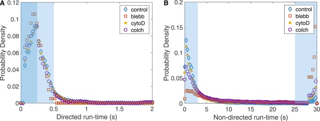
The intracellular environment is a dynamic space filled with various organelles moving in all directions. Included in this diverse group of organelles are vesicles, which are involved in transport of molecular cargo throughout the cell. Vesicles move in either a directed or non-directed fashion, often depending on interactions with cytoskeletal proteins such as microtubules, actin filaments, and molecular motors. How these proteins affect the local fluctuations of vesicles in the cytoplasm is not clear since they have the potential to both facilitate and impede movement. Here we show that vesicle mobility is significantly affected by myosin-II, even though it is not a cargo transport motor. We find that myosin-II activity increases the effective diffusivity of vesicles and its inhibition facilitates longer states of non-directed motion. Our study suggests that altering myosin-II activity in the cytoplasm of cells can modulate the mobility of vesicles, providing a possible mechanism for cells to dynamically tune the cytoplasmic environment in space and time.


Conflict of interest statement

The authors declare no competing interests.

Figures

Figure 1
Figure 1
Trajectories of vesicle motion in fibroblasts. Two examples of raw data for the vesicle positions are shown. The temporal mean squared displacement (tMSD) analysis was used to identify directed and non-directed motion. (A) A trajectory exhibiting short bursts of directed motion (v ~ 500 nm/s) followed by a long period of non-directed motion (~25 s). (B) Another trajectory showing directed motion (v ~ 700 nm/s) and non-directed motion (~8 s). The maximum possible trajectory duration in this study is 30 s due to acquisition length.
Figure 2
Figure 2
Vesicle dynamics does not depend on substrate stiffness. (A) Mean squared displacement (MSD) of vesicle motion in untreated cells indicates no strong dependence on substrate stiffness. Vesicles in all cases show sub-diffusive behavior at shorter timescales transitioning to diffusive behavior at longer timescales. van Hove Correlation (VHC) functions for vesicles undergoing directed motion (B) and non-directed motion (C) show the full distribution of vesicle displacements is not sensitive to substrate stiffness. VHCs calculated for a timescale of 150 ms. Notice that VHCs of both directed and non-directed motion are strongly non-Gaussian, indicating cytoplasmic activity. (Number of experiments/cells/trajectories - 10 kPa: 7/28/1211, 40 kPa: 7/24/3021, glass: 10/31/5605; S.E.M. smaller than symbol size).
Figure 3
Figure 3
Velocity statistics indicate robustness to substrate stiffness and sensitivity to cytoskeletal perturbation. (A) The velocity distribution of directed motion is independent of substrate stiffness. This provides further support, in addition to MSDs and VHCs, that substrate stiffness does not affect vesicle motion in fibroblasts. (B) The velocity autocorrelation function (VACF) quantifies persistence and indicates that vesicle motion is strongly perturbed by blebbistatin, which inactivates myosin-II. The strong negative peak (red squares) indicates vesicle motion is decreased when myosin-II is inactivated. Data shown for cells cultured on 10 kPa substrate.
Figure 4
Figure 4
Directed run-times are robust, but non-directed run-times are sensitive to cytoskeletal perturbation. (A) The directed run-time distribution indicates vesicles spend a similar amount of time undergoing directed motion independent of cytoskeletal perturbation. These durations are relatively short, typically less than 500 ms. (B) Non-directed run-times indicate two timescales of random motion, short (<3 s) and long (>25 s) for all conditions. When cytoskeletal perturbations are introduced, there is a shift in non-directed motion from short to long run-times. Inactivation of myosin-II has the strongest effect, shifting nearly half of vesicles to experience mainly non-directed motion.
Figure 5
Figure 5
Vesicle motion is more sensitive to cytoskeletal perturbation on softer substrates. (A) Vesicle dynamics on glass is not strongly affected by cytoskeletal perturbation. However, the effect becomes more pronounced on (B) 40 kPA gels and (C) 10 kPa gels. Relative to the control vesicle motion, MSD plots suggest colchicine treatment decreases motion and cyto-D increases motion. (Number of experiments/cells/trajectories - glass: control 13/31/6738, blebb 8/19/4498, colch 8/16/2111, cytoD 8/18/3984; 40 kPa: control 10/30/3484, blebb 8/16/2412, colch 8/16/1324, cytoD 8/16/2139; 10 kPa: control 10/27/1855, blebb 8/15/773, colch 8/10/532, cytoD 8/12/797; S.E.M. shown as shaded background).

Similar articles

Cited by

References

    1. Ross JL, Ali MY, Warshaw DM. Cargo transport: molecular motors navigate a complex cytoskeleton. Curr. opinion cell biology. 2008;20:41–47. doi: 10.1016/j.ceb.2007.11.006. - DOI - PMC - PubMed
    1. Caspi A, Granek R, Elbaum M. Diffusion and directed motion in cellular transport. Phys Rev E. 2002;66:011916. doi: 10.1103/PhysRevE.66.011916. - DOI - PubMed
    1. Arcizet D, Meier B, Sackmann E, Rädler JO, Heinrich D. Temporal analysis of active and passive transport in living cells. Phys Rev Lett. 2008;101:248103. doi: 10.1103/PhysRevLett.101.248103. - DOI - PubMed
    1. Ahmed WW, Williams BJ, Silver AM, Saif TA. Measuring nonequilibrium vesicle dynamics in neurons under tension. Lab on a Chip. 2013;13:570–578. doi: 10.1039/c2lc41109a. - DOI - PubMed
    1. Koslover EF, Chan CK, Theriot JA. Cytoplasmic flow and mixing due to deformation of motile cells. Biophys. J. 2017;113:2077–2087. doi: 10.1016/j.bpj.2017.09.009. - DOI - PMC - PubMed

Publication types

LinkOut - more resources