

Bright GFP with subnanosecond fluorescence lifetime
- PMID: 30185895
- PMCID: PMC6125319
- DOI: 10.1038/s41598-018-31687-w
Bright GFP with subnanosecond fluorescence lifetime
Abstract
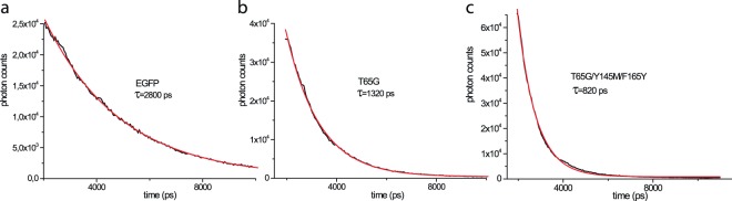
Fluorescence lifetime imaging microscopy (FLIM) measures fluorescence decay rate at every pixel of an image. FLIM can separate probes of the same color but different fluorescence lifetimes (FL), thus it is a promising approach for multiparameter imaging. However, available GFP-like fluorescent proteins (FP) possess a narrow range of FLs (commonly, 2.3-3.5 ns) which limits their applicability for multiparameter FLIM. Here we report a new FP probe showing both subnanosecond fluorescence lifetime and exceptional fluorescence brightness (80% of EGFP). To design this probe we applied semi-rational amino acid substitutions selection. Critical positions (Thr65, Tyr145, Phe165) were altered based on previously reported effect on FL or excited state electron transfer. The resulting EGFP triple mutant, BrUSLEE (Bright Ultimately Short Lifetime Enhanced Emitter), allows for both reliable detection of the probe and recording FL signal clearly distinguishable from that of the spectrally similar commonly used GFPs. We demonstrated high performance of this probe in multiparameter FLIM experiment. We suggest that amino acid substitutions described here lead to a significant shift in radiative and non-radiative excited state processes equilibrium.
Conflict of interest statement
The authors declare no competing interests.
Figures

References
Publication types
MeSH terms
Substances
Grants and funding
- 17-00-00401/Russian Foundation for Basic Research (RFBR)/International
- 17-00-00403/Russian Foundation for Basic Research (RFBR)/International
- 17-00-00401/Russian Foundation for Basic Research (RFBR)/International
- RFMEFI62117X0018/Ministry of Education and Science of the Russian Federation/International
- RFMEFI62117X0018/Ministry of Education and Science of the Russian Federation/International
LinkOut - more resources
Full Text Sources
Other Literature Sources
Miscellaneous
