TiHo-0906: a new feline mammary cancer cell line with molecular, morphological, and immunocytological characteristics of epithelial to mesenchymal transition
- PMID: 30185896
- PMCID: PMC6125410
- DOI: 10.1038/s41598-018-31682-1
TiHo-0906: a new feline mammary cancer cell line with molecular, morphological, and immunocytological characteristics of epithelial to mesenchymal transition
Abstract
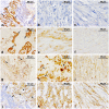
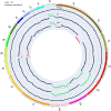
Feline mammary carcinomas (FMCs) with anaplastic and malignant spindle cells histologically resemble the human metaplastic breast carcinoma (hMBC), spindle-cell subtype. hMBCs display epithelial-to-mesenchymal transition (EMT) characteristics. Herein we report the establishment and characterization of a cell line (TiHoCMglAdcar0906; TiHo-0906) exhibiting EMT-like properties derived from an FMC with anaplastic and malignant spindle cells. Copy-number variations (CNVs) by next-generation sequencing and immunohistochemical characteristics of the cell line and the tumour were compared. The absolute qPCR expression of EMT-related markers HMGA2 and CD44 was determined. The growth, migration, and sensitivity to doxorubicin were assessed. TiHo-0906 CNVs affect several genomic regions harbouring known EMT-, breast cancer-, and hMBCs-associated genes as AKT1, GATA3, CCND2, CDK4, ZEB1, KRAS, HMGA2, ESRP1, MTDH, YWHAZ, and MYC. Most of them were located in amplified regions of feline chromosomes (FCAs) B4 and F2. TiHo-0906 cells displayed an epithelial/mesenchymal phenotype, and high HMGA2 and CD44 expression. Growth and migration remained comparable during subculturing. Low-passaged cells were two-fold more resistant to doxorubicin than high-passaged cells (IC50: 99.97 nM, and 41.22 nM, respectively). The TiHo-0906 cell line was derived from a poorly differentiated cellular subpopulation of the tumour consistently displaying EMT traits. The cell line presents excellent opportunities for studying EMT on FMCs.
Conflict of interest statement
The authors declare no competing interests.
Figures


Similar articles
-
Analysis of Copy-Number Variations and Feline Mammary Carcinoma Survival.Sci Rep. 2020 Jan 22;10(1):1003. doi: 10.1038/s41598-020-57942-7. Sci Rep. 2020. PMID: 31969654 Free PMC article.
-
WNT5A signaling impairs breast cancer cell migration and invasion via mechanisms independent of the epithelial-mesenchymal transition.J Exp Clin Cancer Res. 2016 Sep 13;35(1):144. doi: 10.1186/s13046-016-0421-0. J Exp Clin Cancer Res. 2016. PMID: 27623766 Free PMC article.
-
Foxf2 plays a dual role during transforming growth factor beta-induced epithelial to mesenchymal transition by promoting apoptosis yet enabling cell junction dissolution and migration.Breast Cancer Res. 2018 Oct 1;20(1):118. doi: 10.1186/s13058-018-1043-6. Breast Cancer Res. 2018. PMID: 30285803 Free PMC article.
-
Cancer stem-like cells enriched with CD29 and CD44 markers exhibit molecular characteristics with epithelial-mesenchymal transition in squamous cell carcinoma.Arch Dermatol Res. 2013 Jan;305(1):35-47. doi: 10.1007/s00403-012-1260-2. Epub 2012 Jun 28. Arch Dermatol Res. 2013. PMID: 22740085
-
The GRHL2/ZEB Feedback Loop-A Key Axis in the Regulation of EMT in Breast Cancer.J Cell Biochem. 2017 Sep;118(9):2559-2570. doi: 10.1002/jcb.25974. Epub 2017 May 3. J Cell Biochem. 2017. PMID: 28266048
Cited by
-
Characterization of six canine prostate adenocarcinoma and three transitional cell carcinoma cell lines derived from primary tumor tissues as well as metastasis.PLoS One. 2020 Mar 13;15(3):e0230272. doi: 10.1371/journal.pone.0230272. eCollection 2020. PLoS One. 2020. PMID: 32168360 Free PMC article.
-
Effects of hsa-miR-28-5p on Adriamycin Sensitivity in Diffuse Large B-Cell Lymphoma.Evid Based Complement Alternat Med. 2022 Jul 13;2022:4290994. doi: 10.1155/2022/4290994. eCollection 2022. Evid Based Complement Alternat Med. 2022. PMID: 35873635 Free PMC article.
-
Transcription profiling of feline mammary carcinomas and derived cell lines reveals biomarkers and drug targets associated with metabolic and cell cycle pathways.Sci Rep. 2022 Oct 11;12(1):17025. doi: 10.1038/s41598-022-20874-5. Sci Rep. 2022. PMID: 36220861 Free PMC article.
-
Establishment and Characterization of Feline Mammary Tumor Patient-Derived Xenograft Model.Animals (Basel). 2021 Aug 12;11(8):2380. doi: 10.3390/ani11082380. Animals (Basel). 2021. PMID: 34438836 Free PMC article.
-
Analysis of Copy-Number Variations and Feline Mammary Carcinoma Survival.Sci Rep. 2020 Jan 22;10(1):1003. doi: 10.1038/s41598-020-57942-7. Sci Rep. 2020. PMID: 31969654 Free PMC article.
References
-
- Misdorp, W., Else, R., Hellmen, E. & Lipscomb, T. Histological classification of mammary tumors of the dog and cat. (1999).
-
- Ellis, I. et al. In Pathology and Genetics of Tumours of the Breast and Female Organs. WHO Classification of Tumours. (eds Tavassoli, F. A. & Devilee, P.) 37–38 (IARCPress, 2003).
MeSH terms
LinkOut - more resources
Full Text Sources
Other Literature Sources
Medical
Research Materials
Miscellaneous

