. 2018 Sep 5;8(1):13259.
doi: 10.1038/s41598-018-31525-z.

Functional diversity of brain networks supports consciousness and verbal intelligence

Affiliations

Functional diversity of brain networks supports consciousness and verbal intelligence

Lorina Naci et al. Sci Rep. .

Abstract

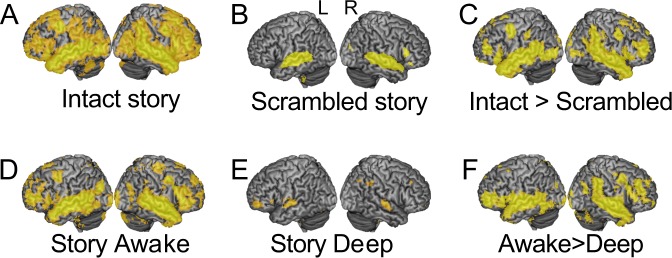
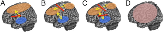
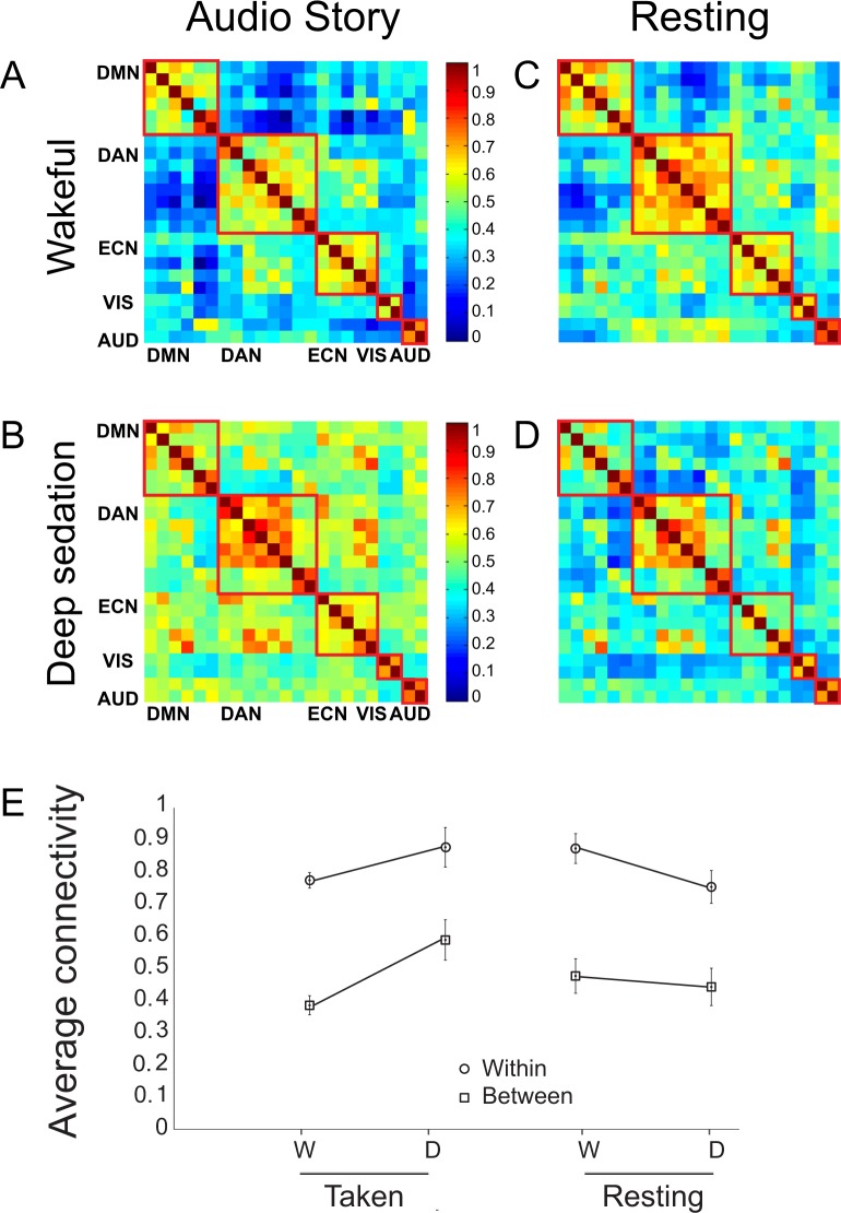
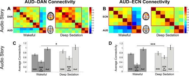
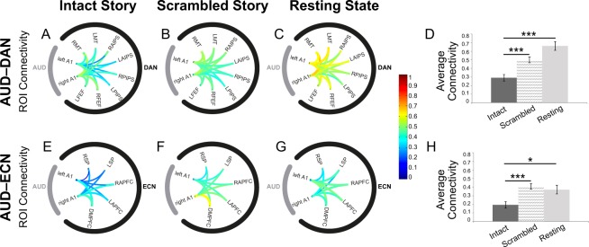
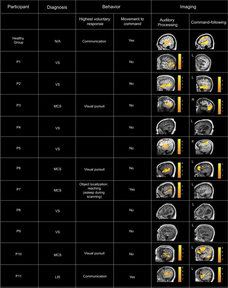
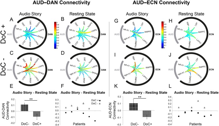
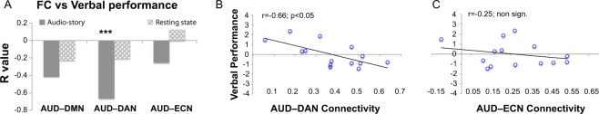
How are the myriad stimuli arriving at our senses transformed into conscious thought? To address this question, in a series of studies, we asked whether a common mechanism underlies loss of information processing in unconscious states across different conditions, which could shed light on the brain mechanisms of conscious cognition. With a novel approach, we brought together for the first time, data from the same paradigm-a highly engaging auditory-only narrative-in three independent domains: anesthesia-induced unconsciousness, unconsciousness after brain injury, and individual differences in intellectual abilities during conscious cognition. During external stimulation in the unconscious state, the functional differentiation between the auditory and fronto-parietal systems decreased significantly relatively to the conscious state. Conversely, we found that stronger functional differentiation between these systems in response to external stimulation predicted higher intellectual abilities during conscious cognition, in particular higher verbal acuity scores in independent cognitive testing battery. These convergent findings suggest that the responsivity of sensory and higher-order brain systems to external stimulation, especially through the diversification of their functional responses is an essential feature of conscious cognition and verbal intelligence.


Conflict of interest statement

The authors declare no competing interests.

Figures

Figure 1
Figure 1
Brain-wide inter-subject correlation of neural activity during the audio story. (A) The audio story elicited significant (p < 0.05; FWE cor) inter-subject correlation across the brain, including frontal and parietal cortex, thought to support executive function. (B) The baseline elicited significant (p < 0.05; FWE cor) inter-subject correlation within primary and association auditory cortex. A small cluster was also observed in right inferior prefrontal cortex. None was observed in dorsal prefrontal and parietal cortex. (C) The audio story elicited significantly (p < 0.05; FWE cor) more inter-subject correlation than the auditory baseline derived from the same stimulus, in parietal, temporal, motor, and dorsal/ventral frontal/prefrontal cortex. A, B, C, adapted with permission from Naci et al.. (D) The audio story elicited significant (p < 0.05; FWE cor) inter-subject correlation across the brain, including frontal and parietal cortex, in the wakeful state of the anesthesia study. (E) In the deep anesthesia state, significant (p < 0.05; FWE cor) inter-subject correlation was limited to the auditory cortex with the exception of two small clusters, one in left prefrontal and the other in right parietal cortex. (F) The audio story elicited significantly (p < 0.05; FWE cor) more cross-subject correlation in the awake than deeply sedated condition bilaterally in temporal, ventral prefrontal and frontal cortex, and further in parietal, motor, and dorsal frontal and prefrontal cortex in the right hemisphere. Warmer colors depict higher t-values of cross-subject correlation. Warmer colors depict higher t-values of inter-subject correlation.
Figure 2
Figure 2
Candidate patterns of connectivity perturbations caused by deep propofol anesthesia. (A) Loss of long-range connectivity between different networks; (B) Loss of long-range connectivity within a specific network, e.g., between frontal and parietal regions; (C) A combination of patterns in A and B; (D) Loss of functional differentiation between different brain networks.
Figure 3
Figure 3
Global within- and between-network functional connectivity perturbations by deep propofol anesthesia (AD) Functional connectivity matrices for five brain networks in the story and resting state conditions, in the wakeful and deep anesthesia states. Each cell represents the correlation of the time-course of one region of interest (ROI) with another, or itself (in the center diagonal). Cells representing correlations of ROIs within each network are delineated by red squares. Warm/cool colors represent high/low correlations, as shown in heat-bar scale. (E) Average connectivity (z values) within- and between- networks in the wakeful (W) and the deep anesthesia (D) states, during the story and resting state conditions. DMN/DAN/ECN/VIS/AUD = Default Mode/Dorsal Attention/Executive Control/Visual/Auditory network.
Figure 4
Figure 4
Perturbations of auditory and fronto-parietal connectivity by deep propofol anesthesia in the audio story condition. Only connectivity between the AUD and DAN/ECN, respectively, was significantly modulated by propofol, showing a significant reduction of functional differentiation between sensory and higher-order networks in deep anesthesia relative to wakefulness. (A,B) Functional connectivity matrices for ROIs comprising the DAN and AUD (A)/ECN and AUD (B) networks in the wakeful and deep anesthesia states of the audio story condition. (C,D) Average connectivity (z-values) within and between the DAN and AUD (C)/ECN and AUD (D), in the wakeful and deep anesthesia states.
Figure 5
Figure 5
Functional connectivity between the auditory and fronto-parietal networks in healthy wakeful individuals, during the audio story and baseline conditions. Connectivity between the auditory and fronto-parietal networks was significantly modulated by the presence of complex meaningful stimuli, with the functional differentiation between the AUD and DAN/ECN increasing significantly in the audio story as compared to the scrambled story and resting state baseline conditions. (AC) Connectivity between the ROIs within the AUD and DAN networks in the intact story (A), scrambled story (B), and resting state (C) baseline. (D) Average AUD–DAN connectivity (z values) for each condition. (EG) Connectivity between the ROIs within the AUD and ECN networks in the intact story (E), scrambled story (F), and resting state (G) baseline. (H) Average AUD–ECN connectivity (z values) for each condition. A1: Primary auditory cortex; LFEF: Left frontal eye field; RFEF: Right frontal eye field; LPIPS: Left posterior IPS; RPIPS: Right posterior IPS; LAIPS Left anterior IPS; RAIPS: Right anterior IPS LMT: Left middle temporal area; RMT: Right middle temporal area; DMPFC: Dorsal medial PFC; LAPFC: Left anterior prefrontal cortex; RAPFC: Right anterior prefrontal cortex LSP: Left superior parietal; RSP: Right superior parietal.
Figure 6
Figure 6
Summary of DoC patients’ clinical and fMRI assessment data. Auditory processing. In the fMRI assessment, three patients clinically diagnosed to be in a VS did not show evidence of auditory processing. The other eight patients who showed evidence of auditory processing, two patients clinically diagnosed as VS did not show evidence of brain-based command-following, and the other six, including two diagnosed as VS, showed evidence of brain-based command-following, and thus, of covert awareness. Command-following. 6/11 patients followed task commands by willfully modulating their brain activity as requested, and thus, provided evidence of conscious awareness. Two of these (P2, P5) presented a CMD profile, or a behavioral diagnosis of VS that was inconsistent with their positive fMRI results. 5/11 patients showed no evidence of willful responses in the fMRI command-following task, and, thus, provided no neuroimaging evidence of awareness. One (P7) showed no neuroimaging evidence of awareness despite an MCS diagnosis, due to falling asleep in the scanner for the entirely of the session (Materials and Methods).
Figure 7
Figure 7
Modulation of auditory to fronto-parietal connectivity by meaningful stimulation in DoC patients. Similarly to healthy individuals, connectivity between the auditory and fronto-parietal networks in DoC+ patients was significantly modulated by the presence of complex meaningful stimuli, with the functional differentiation between the AUD and DAN/ECN increasing significantly in the audio story as compared to the resting state baseline condition. (AD) Connectivity between the ROIs within the AUD and DAN networks, during the audio story and resting state baseline, in the DoC+ (A,B) and DoC− (C,D) patients. (E) Differential averaged AUD–DAN connectivity (z values) for each patient group. (F) Differential averaged AUD–DAN connectivity (z values) for each individual patient. (GJ) Connectivity between the ROIs within the AUD and ECN networks, during the audio story and resting state baseline, in the DoC+ (G,H) and DoC− (I,J) patients. (K) Differential averaged AUD–ECN connectivity (z values) for each patient group. (L) Differential averaged AUD–ECN connectivity (z values) for each individual patient. A1: Primary auditory cortex; LFEF: Left frontal eye field; RFEF: Right frontal eye field; LPIPS: Left posterior IPS; RPIPS: Right posterior IPS; LAIPS Left anterior IPS; RAIPS: Right anterior IPS LMT: Left middle temporal area; RMT: Right middle temporal area; DMPFC: Dorsal medial PFC; LAPFC: Left anterior prefrontal cortex; RAPFC: Right anterior prefrontal cortex LSP: Left superior parietal; RSP: Right superior parietal.
Figure 8
Figure 8
The relationship between brain network connectivity during the audio story and independently-measured cognitive performance. The functional connectivity between AUD and DAN, but not ECN (or DMN, used here as a high-level control network) during the story, and not the resting state baseline condition, was significantly inversely related to verbal performance. (A) Group-averaged correlation between the functional connectivity (FC) of the AUD and the DMN/DAN/ECN networks during the audio story and resting state conditions and verbal performance. (B,C) For each participant, the correlation between their AUD–DAN (B)/AUD–ECN (C) connectivity during the story and their verbal performance is displayed.

Similar articles

Cited by

References

    1. Dehaene S, Changeux JP. Experimental and theoretical approaches to conscious processing. Neuron. 2011;70(2):200–27. doi: 10.1016/j.neuron.2011.03.018. - DOI - PubMed
    1. Dehaene S, Changeux J, Naccache L, Sackur J, Sergent C. Conscious, preconscious, and subliminal processing: a testable taxonomy. Trends In Cognitive Sciences. 2006;10:204–211. doi: 10.1016/j.tics.2006.03.007. - DOI - PubMed
    1. Lamme VAF, Roelfsema PR. The distinct modes of vision offered by feedforward and recurrent processing. Trends Neurosci. 2000;23:571–579. doi: 10.1016/S0166-2236(00)01657-X. - DOI - PubMed
    1. Dehaene S, Charles L, King JR, Marti S. Toward a computational theory of conscious processing. Curr. Opin. Neurobiol. 2014;25:76–84. doi: 10.1016/j.conb.2013.12.005. - DOI - PMC - PubMed
    1. MacDonald A, Naci L, MacDonald P, Owen AM. Anesthesia and neuroimaging: Investigating the neural correlates of unconsciousness. TICS. 2015;19(2):100–107. - PubMed

Publication types

LinkOut - more resources