

MRI Asymmetry Index of Hippocampal Subfields Increases Through the Continuum From the Mild Cognitive Impairment to the Alzheimer's Disease
- PMID: 30186103
- PMCID: PMC6111896
- DOI: 10.3389/fnins.2018.00576
MRI Asymmetry Index of Hippocampal Subfields Increases Through the Continuum From the Mild Cognitive Impairment to the Alzheimer's Disease
Abstract
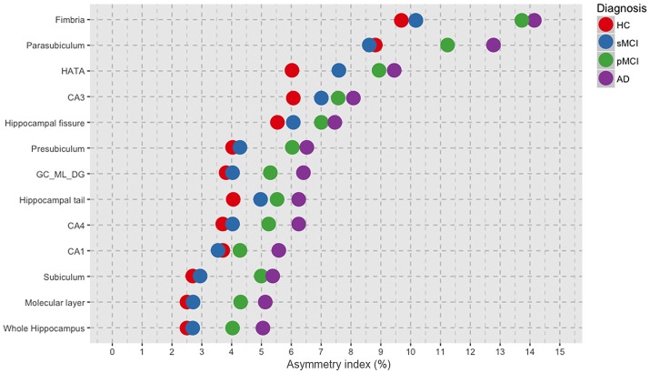
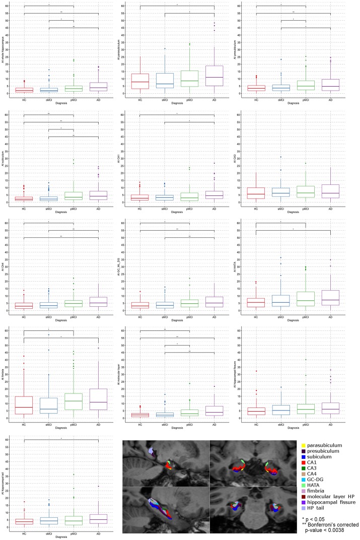
Objective: It is well-known that the hippocampus presents significant asymmetry in Alzheimer's disease (AD) and that difference in volumes between left and right exists and varies with disease progression. However, few works investigated whether the asymmetry degree of subfields of hippocampus changes through the continuum from Mild Cognitive Impairment (MCI) to AD. Thus, aim of the present work was to evaluate the Asymmetry Index (AI) of hippocampal substructures as possible MRI biomarkers of Dementia. Moreover, we aimed to assess whether the subfields presented peculiar differences between left and right hemispheres. We also investigated the relationship between the asymmetry magnitude in hippocampal subfields and the decline of verbal memory as assessed by Rey's auditory verbal learning test (RAVLT). Methods: Four-hundred subjects were selected from ADNI, equally divided into healthy controls (HC), AD, stable MCI (sMCI), and progressive MCI (pMCI). The structural baseline T1s were processed with FreeSurfer 6.0 and volumes of whole hippocampus (WH) and 12 subfields were extracted. The AI was calculated as: (|Left-Right|/(Left+Right))*100. ANCOVA was used for evaluating AI differences between diagnoses, while paired t-test was applied for assessing changes between left and right volumes, separately for each group. Partial correlation was performed for exploring relationship between RAVLT summary scores (Immediate, Learning, Forgetting, Percent Forgetting) and hippocampal substructures AI. The statistical threshold was Bonferroni corrected p < 0.05/13 = 0.0038. Results: We found a general trend of increased degree of asymmetry with increasing severity of diagnosis. Indeed, AD presented the higher magnitude of asymmetry compared with HC, sMCI and pMCI, in the WH (AI mean 5.13 ± 4.29 SD) and in each of its twelve subfields. Moreover, we found in AD a significant negative correlation (r = -0.33, p = 0.00065) between the AI of parasubiculum (mean 12.70 ± 9.59 SD) and the RAVLT Learning score (mean 1.70 ± 1.62 SD). Conclusions: Our findings showed that hippocampal subfields AI varies differently among the four groups HC, sMCI, pMCI, and AD. Moreover, we found-for the first time-that hippocampal substructures had different sub-patterns of lateralization compared with the whole hippocampus. Importantly, the severity in learning rate was correlated with pathological high degree of asymmetry in parasubiculum of AD patients.
Keywords: Alzheimer's disease; Mild Cognitive Impairment; asymmetry index; hippocampal subfields; hippocampus; neuroimaging.
Figures


Similar articles
-
Atrophy asymmetry in hippocampal subfields in patients with Alzheimer's disease and mild cognitive impairment.Exp Brain Res. 2023 Feb;241(2):495-504. doi: 10.1007/s00221-022-06543-z. Epub 2023 Jan 2. Exp Brain Res. 2023. PMID: 36593344
-
Distinct Atrophy Pattern of Hippocampal Subfields in Patients with Progressive and Stable Mild Cognitive Impairment: A Longitudinal MRI Study.J Alzheimers Dis. 2021;79(1):237-247. doi: 10.3233/JAD-200775. J Alzheimers Dis. 2021. PMID: 33252076
-
Choroid plexus volumes and auditory verbal learning scores are associated with conversion from mild cognitive impairment to Alzheimer's disease.Brain Behav. 2024 Jul;14(7):e3611. doi: 10.1002/brb3.3611. Brain Behav. 2024. PMID: 38956818 Free PMC article.
-
Structural magnetic resonance imaging for the early diagnosis of dementia due to Alzheimer's disease in people with mild cognitive impairment.Cochrane Database Syst Rev. 2020 Mar 2;3(3):CD009628. doi: 10.1002/14651858.CD009628.pub2. Cochrane Database Syst Rev. 2020. PMID: 32119112 Free PMC article.
-
A review on Alzheimer's disease classification from normal controls and mild cognitive impairment using structural MR images.J Neurosci Methods. 2023 Jan 15;384:109745. doi: 10.1016/j.jneumeth.2022.109745. Epub 2022 Nov 14. J Neurosci Methods. 2023. PMID: 36395961 Review.
Cited by
-
Editorial: Brain hemispheric specialization and its pathological change revealed by neuroimaging and neuropsychology.Front Neurol. 2022 Nov 4;13:1071148. doi: 10.3389/fneur.2022.1071148. eCollection 2022. Front Neurol. 2022. PMID: 36408509 Free PMC article. No abstract available.
-
Compensation versus deterioration across functional networks in amnestic mild cognitive impairment subtypes.Geroscience. 2025 Apr;47(2):1805-1822. doi: 10.1007/s11357-024-01369-9. Epub 2024 Oct 5. Geroscience. 2025. PMID: 39367933 Free PMC article.
-
Atrophy patterns in hippocampal subregions and their relationship with cognitive function in fibromyalgia patients with mild cognitive impairment.Front Neurosci. 2024 May 23;18:1380121. doi: 10.3389/fnins.2024.1380121. eCollection 2024. Front Neurosci. 2024. PMID: 38846715 Free PMC article.
-
Identifying a Biological Signature of Trauma-Related Neurodegeneration Following Repeated Traumatic Brain Injuries Compared with Healthy Controls.Neurotrauma Rep. 2025 Jul 2;6(1):560-568. doi: 10.1089/neur.2025.0052. eCollection 2025. Neurotrauma Rep. 2025. PMID: 40630648 Free PMC article.
-
A FastSurfer Database for Age-Specific Brain Volumes in Healthy Children: A Tool for Quantifying Localized and Global Brain Volume Alterations in Pediatric Patients.Brain Behav. 2025 Jul;15(7):e70689. doi: 10.1002/brb3.70689. Brain Behav. 2025. PMID: 40685764 Free PMC article.
References
-
- Balthazar M. L., Yasuda C. L., Cendes F., Damasceno B. P. (2010). Learning, retrieval, and recognition are compromised in aMCI and mild AD: are distinct episodic memory processes mediated by the same anatomical structures? J. Int. Neuropsychol. Soc. 16, 205–209. 10.1017/S1355617709990956 - DOI - PubMed
-
- Byrne J. H. (2010). Concise Learning and Memory: The Editor's Selection. London: Academic Press.
Grants and funding
LinkOut - more resources
Full Text Sources
Other Literature Sources