Review
. 2018 Aug 21:11:291.
doi: 10.3389/fnmol.2018.00291. eCollection 2018.

Impaired Glycine Receptor Trafficking in Neurological Diseases

Affiliations
Review

Impaired Glycine Receptor Trafficking in Neurological Diseases

Natascha Schaefer et al. Front Mol Neurosci. .

Abstract

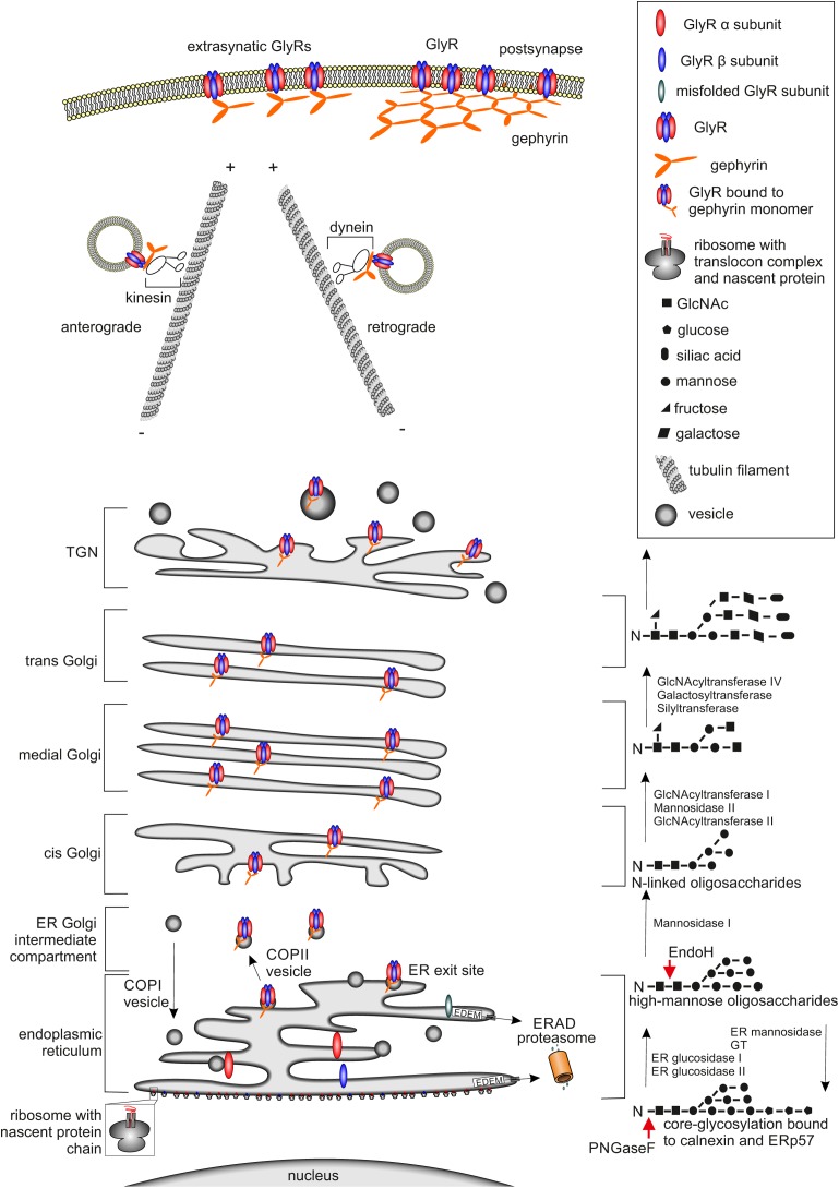
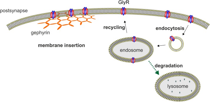
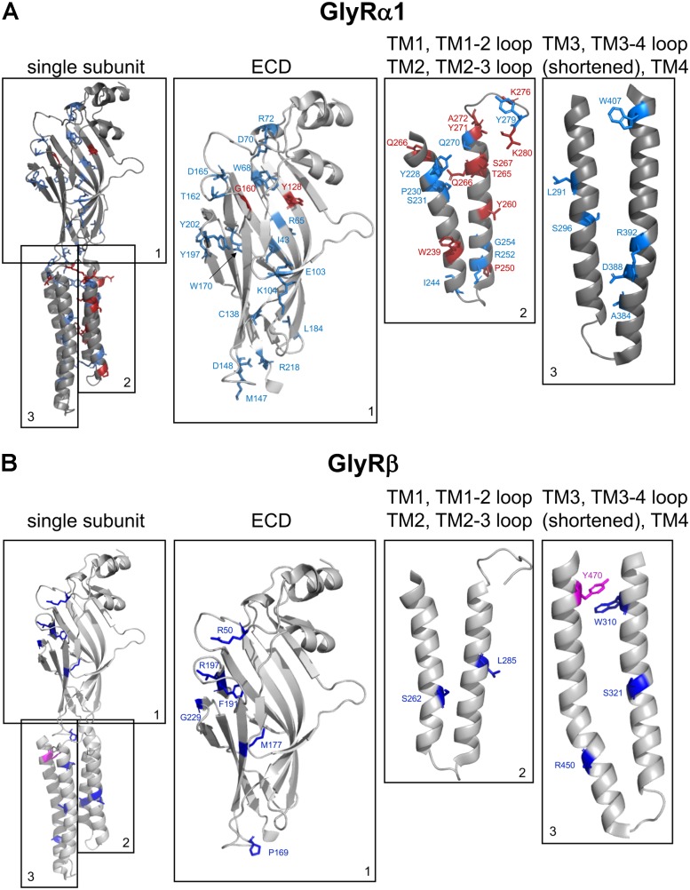
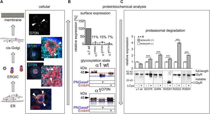
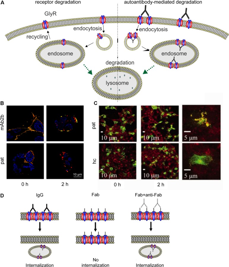
Ionotropic glycine receptors (GlyRs) enable fast synaptic neurotransmission in the adult spinal cord and brainstem. The inhibitory GlyR is a transmembrane glycine-gated chloride channel. The immature GlyR protein undergoes various processing steps, e.g., folding, assembly, and maturation while traveling from the endoplasmic reticulum to and through the Golgi apparatus, where post-translational modifications, e.g., glycosylation occur. The mature receptors are forward transported via microtubules to the cellular surface and inserted into neuronal membranes followed by synaptic clustering. The normal life cycle of a receptor protein includes further processes like internalization, recycling, and degradation. Defects in GlyR life cycle, e.g., impaired protein maturation and degradation have been demonstrated to underlie pathological mechanisms of various neurological diseases. The neurological disorder startle disease is caused by glycinergic dysfunction mainly due to missense mutations in genes encoding GlyR subunits (GLRA1 and GLRB). In vitro studies have shown that most recessive forms of startle disease are associated with impaired receptor biogenesis. Another neurological disease with a phenotype similar to startle disease is a special form of stiff-person syndrome (SPS), which is most probably due to the development of GlyR autoantibodies. Binding of GlyR autoantibodies leads to enhanced receptor internalization. Here we focus on the normal life cycle of GlyRs concentrating on assembly and maturation, receptor trafficking, post-synaptic integration and clustering, and GlyR internalization/recycling/degradation. Furthermore, this review highlights findings on impairment of these processes under disease conditions such as disturbed neuronal ER-Golgi trafficking as the major pathomechanism for recessive forms of human startle disease. In SPS, enhanced receptor internalization upon autoantibody binding to the GlyR has been shown to underlie the human pathology. In addition, we discuss how the existing mouse models of startle disease increased our current knowledge of GlyR trafficking routes and function. This review further illuminates receptor trafficking of GlyR variants originally identified in startle disease patients and explains changes in the life cycle of GlyRs in patients with SPS with respect to structural and functional consequences at the receptor level.

Keywords: autoimmune antibodies; glycine receptor; protein maturation; startle disease; trafficking pathways.


Figures

FIGURE 1
FIGURE 1
Glycine receptor maturation pathway. The GlyR maturation process is depicted. (Left) Schematic cell organelle-structures with important maturation proteins for the GlyRs are shown. (Right) Corresponding linked oligosaccharides to receptors, enzymes enabling glycosylation are labeled. The protein maturation starts with the establishment of single GlyR subunits single oval blue (β subunit) and red (α subunit) structures via translation at the ER. Within the ER, the assembly of single subunits to pentameric GlyR complexes occurs. GlyR complexes are then transferred to the cis-Golgi compartment attached to vesicles where further quality-control mechanisms take place. If GlyRs are misfolded, they will be recycled from the Golgi to the ER possibly by COPI-vesicles and degradation via mechanisms like ERAD leading to the proteasomal pathway occurs. Correctly folded and assembled GlyRs get glycosylated and transported through the secretory Golgi apparatus and trans-Golgi-network (TGN) and shuffled actively via kinesin-anterograde transport along tubulin filaments to the cell surface. The counterpart, retrograde transport of GlyRs is mediated by dynein instead of kinesin, which is responsible for the anterograde transport. At the cellular membrane the GlyR is associated to the scaffold protein gephyrin shown as orange hexagonal-lattice structure.
FIGURE 2
FIGURE 2
Membrane insertion, endocytosis, recycling, and degradation of the GlyR. GlyRs are associated to the hexagonal lattice of the scaffold protein gephyrin when membrane inserted. GlyR degradation occurs by endocytosis as a consequence of ubiquitination followed by lysosomal or lysosomal-like vacuolar degradation. From the endosome, GlyRs can also be recycled and transported back to the cellular surface and transported back to the cellular membrane.
FIGURE 3
FIGURE 3
Structure of a single GlyR subunit including mutations associated with startle disease. Side view onto the principle subunit of GlyR α1 and β based on GlyR α3 (5VDH, Huang et al., 2015). (A) GlyR α1 subunit with marked residues by numbers of the amino acids affected by recessive (blue) and dominant (red) mutations (see also Table 1), parts of the GlyR, e.g., ECD (1); TM1, TM1-2 loop, TM2, and TM2-3 loop (2); TM3, TM3-4 loop (shortened according to Huang et al., 2015) and TM4 (3). (B) Single GlyR β subunit with residues labeled that have been identified as recessive (dark-blue) and dominant (pink) mutations (see also Table 2). The following mutations are not shown due to lack of structural information: residues within the TM3-4 loop – GlyR α1 R316X, G342S and G3475X and GlyR β E24X. Images were made using The PyMOL Molecular Graphics System, Version 1.8 Schrödinger, LLC.
FIGURE 4
FIGURE 4
Biogenesis and trafficking defects of GlyR α1 mutants. (A) GlyR α1 subunits carrying recessive mutations are associated with trafficking defects to the cellular surface. Compartmental analysis of transfected COS7 cells with the GlyR α1 mutant α1D70N showed very few protein dots (marked by white arrow heads) at the cellular surface (labeled before fixation with the α1 specific antibody mAb2b, 1:500). GlyR α1 protein analysis on the ER-ERGIC-cis-Golgi trafficking route determined GlyR α1D70N staining in all compartments analyzed (for ER – calnexin cyan, GlyR α1 red; ERGIC – ERGic53 red, GlyR α1 cyan; cis-Golgi – GM130 red, GlyR α1 cyan). GlyR α1D70N protein accumulation was most pronounced in the ER (large white dots). (B) GlyR protein glycosylation is a pre-requisite for ER exit. The status of protein glycosylation can be determined by digestion with glycosidases Endo H and PNGase F. PNGase F removes all N-linked oligosaccharides from glycoproteins (blue dotted line, lower images). Endo H cuts within the core of high mannose and some hybrid oligosaccharides from N-linked glycoproteins. Once a protein enters the Golgi apparatus and is further glycosylated, the protein gets resistant to Endo H digestion (red dotted line, lower images). A comparison of surface expression between GlyR wild type and mutants can be achieved by biotinylation of surface receptor protein and subsequent binding and elution from streptavidin-beads (upper image). The protein analysis is done normalized to a house keeping protein (e.g., pan-cadherin) and the assumption that the GlyR α1 wild type expression refers to 100%. (C) GlyR α1 mutants are degraded by proteasomal degradation. Using lactacystin (LCys), a proteasomal blocker, GlyR α1 mutant proteins accumulated significantly. The quantification showed significant protein increase following lactacystin treatment for all GlyR α1 mutants but not for the wild type. Images in (A–C) are were modified from Villmann et al. (2009b) and Schaefer et al. (2015). ∗∗∗p < 0.001.
FIGURE 5
FIGURE 5
Native GlyR internalization compared to autoantibody-mediated GlyR internalization. (A) Under native conditions, ubiquitination of cell surface GlyRs initiates receptor degradation by endocytosis followed by lysosomal or lysosomal-like vacuolar degradation (left). Autoantibodies crosslink GlyRs, thus inducing internalization and degradation by endosomal and lysosomal pathways (right). (B) GlyR α1-transfected HEK293 cells were incubated with monoclonal GlyR α1-specific antibody mAb2b or patient serum (pat) and internalization was induced by incubation at 37°C for 0 or 2 h. Patient serum as well as mAb2b were able to induce internalization at 2 h. Red = internalized GlyRs; yellow = membrane-integrated GlyRs; blue = DAPI staining. (C) GlyR α1-EGFP expressing HEK293 cells were incubated with patient serum for 0 or 2 h at 37°C to induce internalization and stained with late endosomal marker LAMP2. Colocalization of both signals is higher in cells incubated with patient serum than with healthy control (hc). Green = GlyR α1-EGFP signal; red = LAMP2 signal (taken with permission from Carvajal-Gonzalez et al., 2014). (D) IgG binding to receptors is able to induce internalization (left), whereas Fab fragments alone cannot elicit receptor internalization (middle). Internalization can re-occur, when Fab fragments and anti-Fab antibodies are incubated together (right), indicating that crosslinking of receptors is required for internalization.

References

    1. Al-Futaisi A. M., Al-Kindi M. N., Al-Mawali A. M., Koul R. L., Al-Adawi S., Al-Yahyaee S. A. (2012). Novel mutation of GLRA1 in Omani families with hyperekplexia and mild mental retardation. Pediatr. Neurol. 46 89–93. 10.1016/j.pediatrneurol.2011.11.008 - DOI - PubMed
    1. Al-Owain M., Colak D., Al-Bakheet A., Al-Hashmi N., Shuaib T., Al-Hemidan A., et al. (2012). Novel mutation in GLRB in a large family with hereditary hyperekplexia. Clin. Genet. 81 479–484. 10.1111/j.1399-0004.2011.01661.x - DOI - PubMed
    1. Arribas-Gonzalez E., de Juan-Sanz J., Aragon C., Lopez-Corcuera B. (2015). Molecular basis of the dominant negative effect of a glycine transporter 2 mutation associated with hyperekplexia. J. Biol. Chem. 290 2150–2165. 10.1074/jbc.M114.587055 - DOI - PMC - PubMed
    1. Atak S., Langlhofer G., Schaefer N., Kessler D., Meiselbach H., Delto C., et al. (2015). Disturbances of ligand potency and enhanced degradation of the human glycine receptor at affected positions G160 and T162 originally identified in patients suffering from hyperekplexia. Front. Mol. Neurosci. 8:79. 10.3389/fnmol.2015.00079 - DOI - PMC - PubMed
    1. Balint B., Bhatia K. P. (2016). Stiff person syndrome and other immune-mediated movement disorders – New insights. Curr. Opin. Neurol. 29 496–506. 10.1097/WCO.0000000000000351 - DOI - PubMed

LinkOut - more resources