. 2018 Aug 21:12:272.
doi: 10.3389/fncel.2018.00272. eCollection 2018.

Costorage of High Molecular Weight Neurotransmitters in Large Dense Core Vesicles of Mammalian Neurons

Affiliations

Costorage of High Molecular Weight Neurotransmitters in Large Dense Core Vesicles of Mammalian Neurons

Adalberto Merighi. Front Cell Neurosci. .

Abstract

It is today widely accepted that several types of high molecular weight (MW) neurotransmitters produced by neurons are synthesized at the cell body, selectively stored within large dense core vesicles (LDCVs) and anterogradely transported to terminals where they elicit their biological role(s). Among these molecules there are neuropeptides and neurotrophic factors, the main focus of this perspective article. I here first provide a brief resume of the state of art on neuronal secretion, with primary emphasis on the molecular composition and mechanism(s) of filling and release of LDCVs. Then, I discuss the perspectives and future directions of research in the field as regarding the synthesis and storage of multiple high MW transmitters in LDCVs and the possibility that a selective sorting of LDCVs occurs along different neuronal processes and/or their branches. I also consider the ongoing discussion that diverse types of neurons may contain LDCVs with different sets of integral proteins or dial in a different fashion with LDCVs containing the same cargo. In addition, I provide original data on the size of LDCVs in rat dorsal root ganglion neurons and their central terminals in the spinal cord after immunogold labeling for calcitonin gene-related peptide (CGRP), neuropeptide K, substance P, neurokinin A or somatostatin. These data corroborate the idea that, similarly to endocrine cells, LDCVs undergo a process of maturation which involves a homotypic fusion followed by a reduction in size and condensation of cargo. They also give support to the conjecture that release at terminals occurs by cavicapture, a process of partial fusion of the vesicle with the axolemma, accompanied by depletion of cargo and diminution of size.

Keywords: co-localization; co-storage; coexistence; large granular vesicles; neuropeptide; neurotransmission; release; small synaptic vesicles.


Figures

Figure 1
Figure 1
Storage of neuropeptides in rat dorsal root ganglion (DRG) neurons. (A,B) Primary afferent terminals in the spinal cord dorsal horn after triple immunogold labeling with antibodies against glutamate (5 nm gold particles), calcitonin gene-related peptide (CGRP; 10 nm gold particles indicated by the yellow arrows in the insert) and substance P (20 nm gold particles indicated by the red arrow heads in the insert). For details on antibodies and labeling methods see Merighi et al. (1991). The two rectangles in (A) are shown at higher magnifications in the inserts. (C) A particular of an axon terminal in the rat dorsal horn after slice incubation in 75 mM K+ to stimulate LDCVs’ exocytosis. Tissue has been processed with the Tannic Acid Ringer Incubation (TARI)-method (Buma et al., 1984). Fixation with tannic acid prior to conventional glutaraldehyde + osmium post-fixation powerfully intensifies the electron density of the substances secreted into the extracellular space. As the cargo of LDCVs is immediately fixed by tannic acid once there, it remains in close apposition to the terminal membrane that appears thicker than after conventional transmission electron microscope (TEM) fixation. Note that three LDCVs in proximity of the axolemma (arrows) display a very electrondense matrix, whereas two other vesicles (arrow heads) display some dissolution of their membranes and are lighter. These images may represent different stages in the process of cavicapture by which neuropeptides may be released at terminals (see also Figure 2). (D,E) Diameter (mean ± SD) of unlabeled, single- and double-labeled LDCVs in the neuronal cell body (D) and central terminals (E) of rat DRG neurons. Note the increase in size of double-labeled LDCVs compared to unlabeled or single-labeled LDCVs in DRGs, likely as a consequence of new cargo addition. Note also that in terminals unlabeled vesicles are smaller than single- and/or double-labeled LDCVs, which, instead, display similar sizes. This observation supports the idea that unlabeled vesicles in terminals may have been depleted of their cargo after cavicapture. Statistics was performed with the GraphPad Prism 7 software. Normality was assessed using the D’Agostino & Pearson normality test. Means were compared using the Kruskal-Wallis non-parametric test followed by Dunn’s multiple comparison. # LDCVs: 346 (cell body), 523 (terminals). ***P = 0.0004; ****P < 0.0001 (two-tailed). Abbreviations: d = dendrite; LDCVs = large dense core vesicles; SSVs = small clear core vesicles. Bars: (A,B) = 200 nm; (C) = 100 nm.
Figure 2
Figure 2
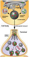
Schematic representation of the process of assembly, filling and release of LDCVs as extrapolated from immunogold staining studies on rat primary sensory neurons in DRGs. For simplicity, only the synthesis of two neuropeptides/proteins (red and green spheres) is depicted. The sizes of immature and mature LDCVs containing no cargo (negative after immunogold labeling), only one of the two molecules (single-labeled) or both molecules (double-labeled) are represented in accordance with the quantitative data reported in Figures 1D,E. In the example, immature LDCVs budding from the trans-golgi network (TGN) contain either one or the other neuropeptide and undergo a process of homotypic fusion to give rise to a larger LDCV. This vesicle stores both peptides, but still may be regarded as immature on the basis of its very large size. It subsequently undergoes a process of condensation with a reduction in size that is completed once the axon terminal is reached by anterograde transport. In terminals, LDCVs are smaller and very likely undergo a process of cavicapture to release their cargo (see also the insert #1 of Figures 1A,C). This process may, in theory, yield to a selective: (a) or a non-selective (b) release of the co-stored neuropeptides. Irrespectively of this possibility, individual LDCVs may endure several cycles of cavicapture until they are fully depleted of their cargo.Quantitative analysis (Figure 1E) demonstrates that empty (unlabeled) LDCVs are smaller than those containing only one (and thus single-labeled) of the two co-stored peptides. To make the figure easier, LDCVs are represented without their outer membrane, which is instead clearly visible in tissues subjected to fixation with tannic acid (Figure 1C). For the same reason, SSVs are not rendered in the terminal.

References

    1. Amara S. G., Arriza J. L., Leff S. E., Swanson L. W., Evans R. M., Rosenfeld M. G. (1985). Expression in brain of a messenger RNA encoding a novel neuropeptide homologous to calcitonin gene-related peptide. Science 229, 1094–1097. 10.1126/science.2994212 - DOI - PubMed
    1. Bartolomucci A., Possenti R., Mahata S. K., Fischer-Colbrie R., Loh Y. P., Salton S. R. (2011). The extended granin family: structure, function, and biomedical implications. Endocr. Rev. 32, 755–797. 10.1210/er.2010-0027 - DOI - PMC - PubMed
    1. Buma P., Roubos E. W., Buijs R. M. (1984). Ultrastructural demonstration of exocytosis of neural, neuroendocrine and endocrine secretions with an in vitro tannic acid (TARI-) method. Histochemistry 80, 247–256. 10.1007/bf00495773 - DOI - PubMed
    1. Cattin-Ortolá J., Topalidou I., Dosey A., Merz A. J., Ailion M. (2017). The dense-core vesicle maturation protein CCCP-1 binds RAB-2 and membranes through its C-terminal domain. Traffic 18, 720–732. 10.1111/tra.12507 - DOI - PMC - PubMed
    1. Cavolo S. L., Bulgari D., Deitcher D. L., Levitan E. S. (2016). activity induces Fmr1-sensitive synaptic capture of anterograde circulating neuropeptide vesicles. J. Neurosci. 36, 11781–11787. 10.1523/JNEUROSCI.2212-16.2016 - DOI - PMC - PubMed