

Assessment of Wound-Healing Properties of Medicinal Plants: The Case of Phyllanthus muellerianus
- PMID: 30186175
- PMCID: PMC6111538
- DOI: 10.3389/fphar.2018.00945
Assessment of Wound-Healing Properties of Medicinal Plants: The Case of Phyllanthus muellerianus
Abstract
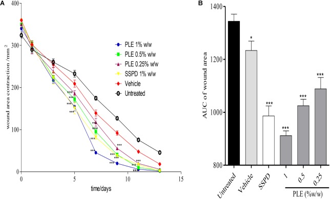
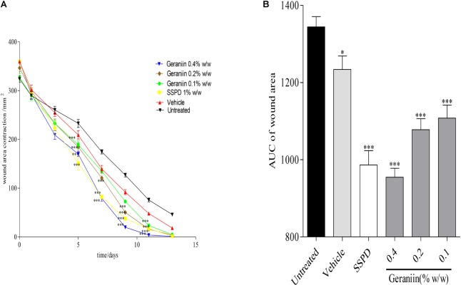
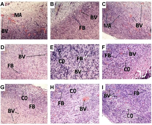
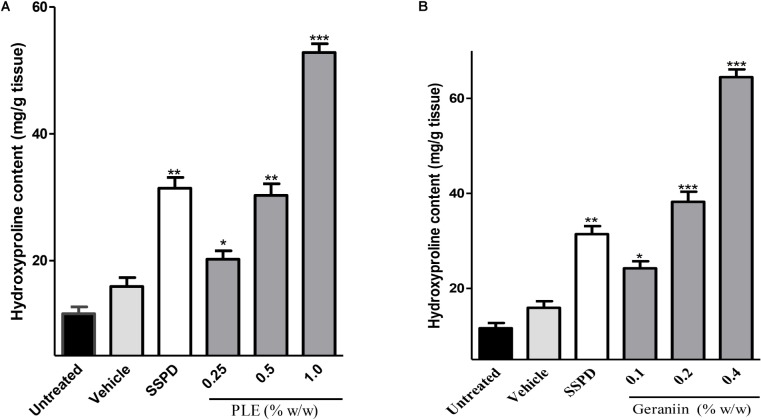
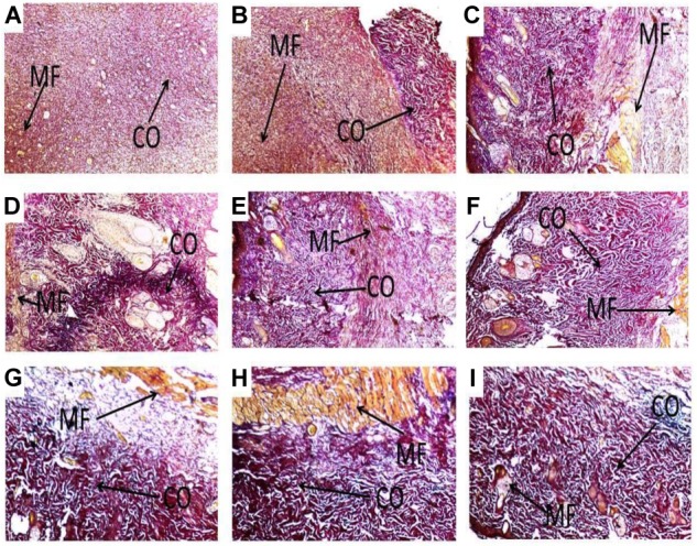
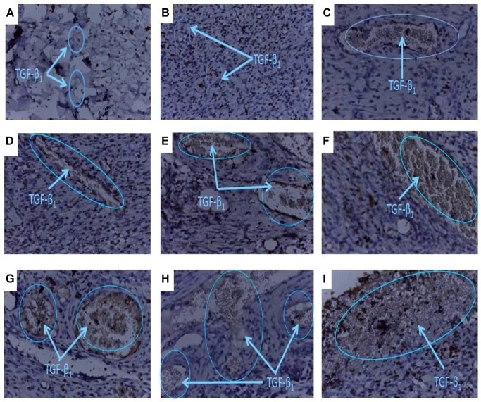
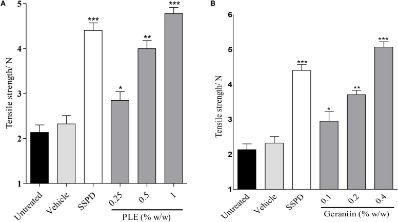
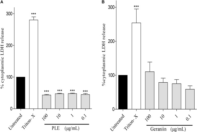
Phyllanthus muellerianus (Family Euphorbiaceae) is a shrub, which is widely distributed in West Africa and employed traditionally as a wound-healing agent especially in Ghana. The aim of the study was to determine the in vivo wound-healing activity of aqueous aerial part extract of P. muellerianus (PLE) and its major isolate, geraniin. Excision and incision wound models were used to determine the wound-healing activity. Wounds were treated with PLE (0.25, 0.5, and 1% w/w) and geraniin (0.1, 0.2, and 0.4% w/w) aqueous creams. PLE and geraniin significantly (p < 0.001) increased wound contraction rate and hydroxyproline production compared to untreated wounds. Histological studies of wound tissues showed high levels of fibroblasts and increased collagen content and cross-linking in PLE and geraniin-treated wound tissues. Immuno-histochemical investigations revealed high levels of TGF-β1 in PLE and geraniin-treated wound tissues compared to the untreated wound tissues. Tensile strength of incised wounds was significantly (p < 0.05) high in PLE and geraniin-treated wounds. PLE (0.1-100 μg/mL) significantly (p < 0.001) reduce LDH release from HaCaT-keratinocytes compared to the untreated cells. PLE and geraniin possess wound healing and cytoprotective effect.
Keywords: Phyllanthus muellerianus; TGF-β1; collagen; cytotoxicity; excision model; geraniin; hydroxyproline; incision model.
Figures
Similar articles
-
Anti-inflammatory activity of aqueous leaf extract of Phyllanthus muellerianus (Kuntze) Exell. and its major constituent, geraniin.J Ethnopharmacol. 2016 Jul 1;187:17-27. doi: 10.1016/j.jep.2016.04.020. Epub 2016 Apr 18. J Ethnopharmacol. 2016. PMID: 27103113
-
Ellagitannins from Phyllanthus muellerianus (Kuntze) Exell.: Geraniin and furosin stimulate cellular activity, differentiation and collagen synthesis of human skin keratinocytes and dermal fibroblasts.Phytomedicine. 2011 May 15;18(7):617-24. doi: 10.1016/j.phymed.2010.08.020. Epub 2010 Oct 30. Phytomedicine. 2011. PMID: 21036574
-
An ethnopharmacological survey and in vitro confirmation of ethnopharmacological use of medicinal plants used for wound healing in Bosomtwi-Atwima-Kwanwoma area, Ghana.J Ethnopharmacol. 2009 Sep 25;125(3):393-403. doi: 10.1016/j.jep.2009.07.024. Epub 2009 Jul 25. J Ethnopharmacol. 2009. PMID: 19635544
-
An isobolographic analysis of the anti-nociceptive effect of geraniin in combination with morphine or diclofenac.J Basic Clin Physiol Pharmacol. 2018 Mar 28;29(2):201-209. doi: 10.1515/jbcpp-2017-0031. J Basic Clin Physiol Pharmacol. 2018. PMID: 28988222
-
Review: African medicinal plants with wound healing properties.J Ethnopharmacol. 2016 Jan 11;177:85-100. doi: 10.1016/j.jep.2015.11.008. Epub 2015 Nov 5. J Ethnopharmacol. 2016. PMID: 26549271 Review.
Cited by
-
Ethnobotanical uses and phytochemical, biological, and toxicological profiles of Datura metel L.: A review.Curr Res Toxicol. 2023 May 13;4:100106. doi: 10.1016/j.crtox.2023.100106. eCollection 2023. Curr Res Toxicol. 2023. PMID: 37228329 Free PMC article.
-
Phytochemical, anti-inflammatory and analgesic properties of stembark extract and fractions of Lonchocarpus sericeus Poir. (Papilionaceae) in albino mice.Avicenna J Phytomed. 2020 Nov-Dec;10(6):584-593. Avicenna J Phytomed. 2020. PMID: 33299815 Free PMC article.
-
Fluoxetine Ecofriendly Nanoemulsion Enhances Wound Healing in Diabetic Rats: In Vivo Efficacy Assessment.Pharmaceutics. 2022 May 26;14(6):1133. doi: 10.3390/pharmaceutics14061133. Pharmaceutics. 2022. Retraction in: Pharmaceutics. 2024 Jan 23;16(2):157. doi: 10.3390/pharmaceutics16020157. PMID: 35745706 Free PMC article. Retracted.
-
Electrospun nanofibers synthesized from polymers incorporated with bioactive compounds for wound healing.J Nanobiotechnology. 2024 Apr 27;22(1):211. doi: 10.1186/s12951-024-02491-8. J Nanobiotechnology. 2024. PMID: 38678271 Free PMC article. Review.
-
Effects of Methanolic Extract Based-Gel From Saudi Pomegranate Peels With Enhanced Healing Potential on Excision Wounds in Diabetic Rats.Front Pharmacol. 2021 May 28;12:704503. doi: 10.3389/fphar.2021.704503. eCollection 2021. Front Pharmacol. 2021. PMID: 34122120 Free PMC article.
References
-
- Adesida G. A., Girgis P., Taylor D. A. H. (1972). Friedelin derivatives from Phyllanthus muellerianus. Phytochemistry 11 851–852. 10.1016/0031-9422(72)80070-0 - DOI
-
- Agyare C., Asase A., Lechtenberg M., Niehues M., Deters A., Hensel A. (2009). An ethnopharmacological survey and in vitro confirmation of ethnopharmacological use of medicinal plants used for wound healing in Bosomtwi-Atwima-Kwanwoma area, Ghana. J. Ethnopharmacol. 125 393–403. 10.1016/j.jep.2009.07.024 - DOI - PubMed
-
- Agyare C., Lechtenberg M., Deters A., Petereit F., Hensel A. (2011). Ellagitannins from Phyllanthus muellerianus (Kuntze) exell: geraniin and furosin stimulate cellular activity, differentiation and collagen synthesis of human skin keratinocytes and dermal fibroblasts. Phytomedicine 18 617–624. 10.1016/j.phymed.2010.08.020 - DOI - PubMed
-
- Benbow M. (2011). Debridement: wound bed preparation. J. Community Nurs. 25 18–23.
LinkOut - more resources
Full Text Sources
Other Literature Sources