Review
. 2018 Aug 21:9:1517.
doi: 10.3389/fpsyg.2018.01517. eCollection 2018.

Inhibitory Pathways for Processing the Temporal Structure of Sensory Signals in the Insect Brain

Affiliations
Review

Inhibitory Pathways for Processing the Temporal Structure of Sensory Signals in the Insect Brain

Hiroyuki Ai et al. Front Psychol. .

Abstract

Insects have acquired excellent sensory information processing abilities in the process of evolution. In addition, insects have developed communication schemes based on the temporal patterns of specific sensory signals. For instance, male moths approach a female by detecting the spatiotemporal pattern of a pheromone plume released by the female. Male crickets attract a conspecific female as a mating partner using calling songs with species-specific temporal patterns. The dance communication of honeybees relies on a unique temporal pattern of vibration caused by wingbeats during the dance. Underlying these behaviors, neural circuits involving inhibitory connections play a critical common role in processing the exact timing of the signals in the primary sensory centers of the brain. Here, we discuss common mechanisms for processing the temporal patterns of sensory signals in the insect brain.

Keywords: cricket; disinhibition; duration coding; honeybee; moth; postinhibitory rebound; temporal structure; waggle dance.


Figures
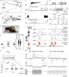
FIGURE 1
FIGURE 1
Insect communication signals and physiologies of critical interneurons involved in temporal processing of sensory signals. (A) Simulated flight path of a moth (winding curve) in a pheromone plume (area enclosed by solid straight lines). Repetitive exposure to sex pheromones in the plume is necessary for sustained upwind flight (from left to right) toward a pheromone source. When the animals lose the pheromonal stimulus, they cease to make upwind progress and instead begin casting. (B) The structure of a plume in turbulent flow from right to left. The shaded area represents the projection of the conical average plume. (C–F) Responses of key neurons in the moth odor processing circuit (see Figure 2A for a circuit diagram). (C) Depolarization-induced firing in the local interneuron 1 (LN1, top) led to hyperpolarization and suppression of firing in the projection neuron (PN, bottom). The PN suppression closely followed the onset of current injection in the LN1 (arrow), and spiking in the PN resumed immediately upon repolarization of the LN1. (D) Hyperpolarizing current injected into an LN1 caused an abrupt suppression of firing of the LN1, and this resulted in depolarization and firing in the PN. This firing in the PN occurred only during LN1 hyperpolarization. (E) Intracellular records from the LN1 and the PN during sex pheromone stimulation (between the two dashed lines). (F) Intracellular records from a local interneuron 2 (LN2, top) and a PN (bottom) responding to brief electrical stimulation of the ipsilateral antennal nerve (asterisks, left). PNs show fast inhibitory postsynaptic potentials (IPSPs; I1 shown left of PN), which disappeared when the gamma-aminobutyric acid (GABA)-receptor blocker bicuculline was applied, resulting in increased variability in the timing of evoked spikes (right of PN). (G) Male crickets produce a calling song by rubbing both forewings together. (H) Audio signal of the calling song in the Mediterranean field cricket (Gryllus bimaculatus). Females are selectively attracted to the pulse pattern of the conspecific calling song. Each chirp has a temporal structure with a fixed pulse period (PP), consisting of pulse duration (PD) and interpulse interval (IPI). (I) Neural network for detecting the temporal (structure of the male cricket calling song. (J) Intracellular membrane potential records of critical interneurons in cricket auditory processing. A postinhibitory rebound (PIR, indicated by asterisks) excitation plays a critical role in song detection. (K) Moving trajectory of a honeybee during the waggle dance. The dance consists of a waggle phase (WP) and a return phase. The distance to the flower source is encoded as the duration of the WP of the dance. (L) Thoracic vibration velocities recorded during the WP. Intermittent vibration pulses occur with a constant PD of about 16 ms and a PP of about 33 ms. (M) Intracellular records of dorsal lobe interneurons 1 (DL-Int-1, middle) and 2 (DL-Int-2, top) in the primary auditory center of the honeybee (see Figure 2E for a circuit diagram) in response to vibratory mechanical stimulation to an antenna (bottom). Left: When the PPs are shorter than 50 ms, the DL-Int-1 receives strong inhibition that allows no spikes during the pulse trains and exhibits a PIR excitation (arrowheads) upon the offset of the pulse train. DL-Int-2 exhibits elevated spiking activity during stimulation. Right: DL-Int-1 shows spikes (asterisks) intermittently during the IPI phase when the PP of the stimulus is longer than 50 ms. Under this condition, the DL-Int-2 often shows a lack of spikes with remarkable IPSPs (dots). Modified from Kaissling (1997) for A; Celani et al. (2014) for B; Christensen et al. (1993) for C, D, and E; Christensen et al. (1998) for F; Hedwig (2016) for H; Schöneich et al. (2015) for I and J; Hrncir et al. (2011) for K and L, with the permission of Birkhäuser Verlag for A; Springer Nature for C, D, and E; The Society for Neuroscience for F; The American Association for the Advancement of Science (AAAS) for I and J; and The Company of Biologists for K and L.)
FIGURE 2
FIGURE 2
Comparison of the neural circuits and response patterns of the moth (A,B), cricket (C,D), and honeybee (E,F). A, C, and E: putative neural circuits for processing the temporal structure of sensory signals. B, D, and F: response of each neuron in these circuits. Disinhibition is found in the moth from LN1 to PN (B) and in the honeybee from DL-Int-1 to DL-Int-2 (F). In the moth, LN2 induces a fast IPSP (I1; thin arrow) for phase-locking the timing of the PN spikes. PIR (thick arrows) excitation may plays a critical role in detecting cricket song IPIs (D) and also in detecting the end of the waggle phase in the honeybee (F).

Similar articles

Cited by

References

    1. Ai H., Kai K., Kumaraswamy A., Ikeno H., Wachtler T. (2017). Interneurons in the honeybee primary auditory center responding to waggle dance-like vibration pulses. J. Neurosci. 37 10624–10635. 10.1523/JNEUROSCI.0044-17.2017 - DOI - PMC - PubMed
    1. Ai H., Nishino H., Itoh T. (2007). Topographic organization of sensory afferents of Johnston’s organ in the honeybee brain. J. Comp. Neurol. 502 1030–1046. 10.1002/cne.21341 - DOI - PubMed
    1. Alluri R. K., Rose G. J., Hanson J. L. (2016). Phasic, suprathreshold excitation and sustained inhibition underlie neuronal selectivity for short-duration sounds. Proc. Natl. Acad. Sci. U.S.A. 113 E1927–E1935. 10.1073/pnas.1520971113 - DOI - PMC - PubMed
    1. Baker T. C., Willis M. A., Haynes K. F., Phelan P. L. (1985). A pulsed cloud of pheromone elicits upwind flight in male moths. Physiol. Entomol. 10 257–265. 10.1111/j.1365-3032.1985.tb00045.x - DOI
    1. Bazhenov M., Stopfer M., Rabinovich M., Abarbanel H. D. I., Sejnowski T., Laurent G. (2001). Model of cellular and network mechanisms for odor-evoked temporal patterning in the locust antennal lobe. Neuron 30 569–581. 10.1016/S0896-6273(01)00286-0 - DOI - PMC - PubMed

LinkOut - more resources