

Primary Lung Dendritic Cell Cultures to Assess Efficacy of Spectinamide-1599 Against Intracellular Mycobacterium tuberculosis
- PMID: 30186246
- PMCID: PMC6110900
- DOI: 10.3389/fmicb.2018.01895
Primary Lung Dendritic Cell Cultures to Assess Efficacy of Spectinamide-1599 Against Intracellular Mycobacterium tuberculosis
Abstract
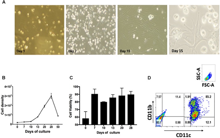
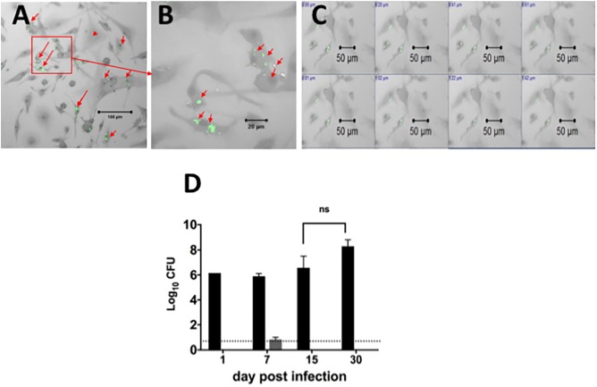
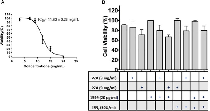
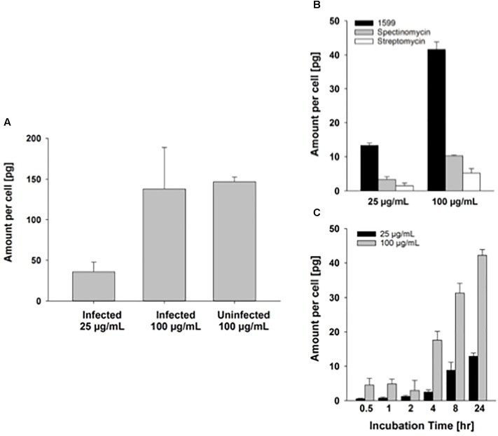
There is an urgent need to treat tuberculosis (TB) quickly, effectively and without side effects. Mycobacterium tuberculosis (Mtb), the causative organism of TB, can survive for long periods of time within macrophages and dendritic cells and these intracellular bacilli are difficult to eliminate with current drug regimens. It is well established that Mtb responds differentially to drug treatment depending on its extracellular and intracellular location and replicative state. In this study, we isolated and cultured lung derived dendritic cells to be used as a screening system for drug efficacy against intracellular mycobacteria. Using mono- or combination drug treatments, we studied the action of spectinamide-1599 and pyrazinamide (antibiotics targeting slow-growing bacilli) in killing bacilli located within lung derived dendritic cells. Furthermore, because IFN-γ is an essential cytokine produced in response to Mtb infection and present during TB chemotherapy, we also assessed the efficacy of these drugs in the presence and absence of IFN-γ. Our results demonstrated that monotherapy with either spectinamide-1599 or pyrazinamide can reduce the intracellular bacterial burden by more than 99.9%. Even more impressive is that when TB infected lung derived dendritic cells are treated with spectinamide-1599 and pyrazinamide in combination with IFN-γ a strong synergistic effect was observed, which reduced the intracellular burden below the limit of detection. We concluded that IFN-γ activation of lung derived dendritic cells is essential for synergy between spectinamide-1599 and pyrazinamide.
Keywords: GM-CSF; Mycobacterium-tuberculosis; dendritic cells; interferon; intracellular; lung; pyrazinamide; spectinamides.
Figures

Similar articles
-
Preclinical Evaluation of Inhalational Spectinamide-1599 Therapy against Tuberculosis.ACS Infect Dis. 2021 Oct 8;7(10):2850-2863. doi: 10.1021/acsinfecdis.1c00213. Epub 2021 Sep 21. ACS Infect Dis. 2021. PMID: 34546724 Free PMC article.
-
Calcitriol enhances pyrazinamide treatment of murine tuberculosis.Chin Med J (Engl). 2019 Sep 5;132(17):2089-2095. doi: 10.1097/CM9.0000000000000394. Chin Med J (Engl). 2019. PMID: 31425356 Free PMC article.
-
The introduction of mesenchymal stromal cells induces different immunological responses in the lungs of healthy and M. tuberculosis infected mice.PLoS One. 2017 Jun 8;12(6):e0178983. doi: 10.1371/journal.pone.0178983. eCollection 2017. PLoS One. 2017. PMID: 28594940 Free PMC article.
-
[Development of antituberculous drugs: current status and future prospects].Kekkaku. 2006 Dec;81(12):753-74. Kekkaku. 2006. PMID: 17240921 Review. Japanese.
-
Pyrazinamide resistance in Mycobacterium tuberculosis: Review and update.Adv Med Sci. 2016 Mar;61(1):63-71. doi: 10.1016/j.advms.2015.09.007. Epub 2015 Oct 1. Adv Med Sci. 2016. PMID: 26521205 Review.
Cited by
-
Dynamic time-kill curve characterization of spectinamide antibiotics 1445 and 1599 for the treatment of tuberculosis.Eur J Pharm Sci. 2019 Jan 15;127:233-239. doi: 10.1016/j.ejps.2018.11.006. Epub 2018 Nov 9. Eur J Pharm Sci. 2019. PMID: 30419293 Free PMC article.
-
Comparative pharmacokinetics of spectinamide 1599 after subcutaneous and intrapulmonary aerosol administration in mice.Tuberculosis (Edinb). 2019 Jan;114:119-122. doi: 10.1016/j.tube.2018.12.006. Epub 2018 Dec 31. Tuberculosis (Edinb). 2019. PMID: 30711150 Free PMC article.
-
Nano-Delivery System of Ethanolic Extract of Propolis Targeting Mycobacterium tuberculosis via Aptamer-Modified-Niosomes.Nanomaterials (Basel). 2023 Jan 8;13(2):269. doi: 10.3390/nano13020269. Nanomaterials (Basel). 2023. PMID: 36678022 Free PMC article.
-
New findings on the incidence and management of CNS adverse reactions in ALK-positive NSCLC with lorlatinib treatment.Discov Oncol. 2024 Sep 13;15(1):444. doi: 10.1007/s12672-024-01339-9. Discov Oncol. 2024. PMID: 39271557 Free PMC article.
-
Development of a Minimalistic Physiologically Based Pharmacokinetic (mPBPK) Model for the Preclinical Development of Spectinamide Antibiotics.Pharmaceutics. 2023 Jun 17;15(6):1759. doi: 10.3390/pharmaceutics15061759. Pharmaceutics. 2023. PMID: 37376207 Free PMC article.
References
-
- Acocella G., Carlone N. A., Cuffini A. M., Cavallo G. (1985). The penetration of rifampicin, pyrazinamide, and pyrazinoic acid into mouse macrophages. Am. Rev. Respir. Dis. 132 1268–1273. - PubMed
-
- Aljayyoussi G., Jenkins V. A., Sharma R., Ardrey A., Donnellan S., Ward S. A., et al. (2017). Pharmacokinetic-pharmacodynamic modelling of intracellular Mycobacterium tuberculosis growth and kill rates is predictive of clinical treatment duration. Sci. Rep. 7:502. 10.1038/s41598-017-00529-6 - DOI - PMC - PubMed
Grants and funding
LinkOut - more resources
Full Text Sources
Other Literature Sources