Review
. 2018 Aug 21:9:1210.
doi: 10.3389/fpls.2018.01210. eCollection 2018.

New Insights Into Wall Polysaccharide O-Acetylation

Affiliations
Review

New Insights Into Wall Polysaccharide O-Acetylation

Markus Pauly et al. Front Plant Sci. .

Abstract

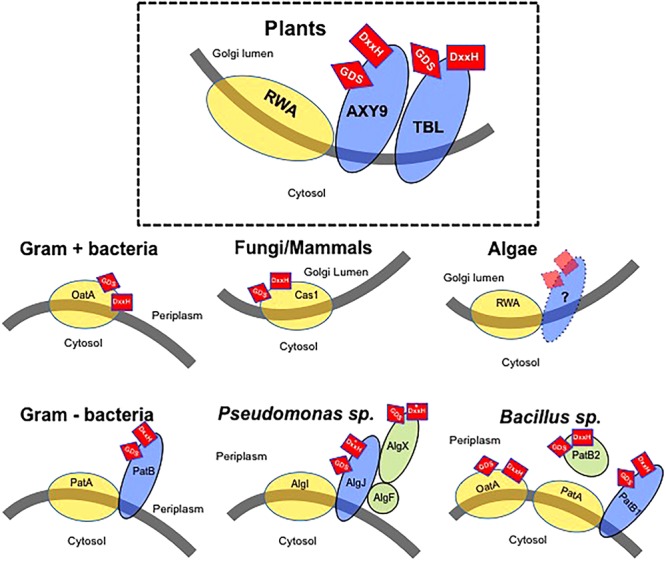
The extracellular matrix of plants, algae, bacteria, fungi, and some archaea consist of a semipermeable composite containing polysaccharides. Many of these polysaccharides are O-acetylated imparting important physiochemical properties to the polymers. The position and degree of O-acetylation is genetically determined and varies between organisms, cell types, and developmental stages. Despite the importance of wall polysaccharide O-acetylation, only recently progress has been made to elucidate the molecular mechanism of O-acetylation. In plants, three protein families are involved in the transfer of the acetyl substituents to the various polysaccharides. In other organisms, this mechanism seems to be conserved, although the number of required components varies. In this review, we provide an update on the latest advances on plant polysaccharide O-acetylation and related information from other wall polysaccharide O-acetylating organisms such as bacteria and fungi. The biotechnological impact of understanding wall polysaccharide O-acetylation ranges from the design of novel drugs against human pathogenic bacteria to the development of improved lignocellulosic feedstocks for biofuel production.

Keywords: O-acetylation; biosynthesis; cell wall; mechanism; polysaccharides.


Figures

FIGURE 1
FIGURE 1
Model of the wall polysaccharides O-acetylation mechanism in various organisms. Gray line – Membrane with indication of cellular location of both sides of the membrane. Yellow circles – Proteins consisting of multitransmembrane domains possibly involved in the translocation of an acetyl-moiety. Blue ovals – Membrane anchored proteins with a single-transmembrane domain. Green circles – Soluble proteins associated with O-acetylation. Red squares – Presence of GDS and DxxH sequences thought to be required for enzymatic activity. The location of these sequences (side of the membrane) is indicated by their location on the protein(s). Asterisk indicates a variation of the consensus DxxH motif (Baker et al., 2014).
FIGURE 2
FIGURE 2
Phylogenetic tree of AXY9, TBL, and RWA proteins. Likelihood tree of AXY9 (A), TBL29 (B), and RWA2 (C) protein homologs constructed from sequence alignment of selected species. Green: embryophytes (Arabidopsis thaliana and Populus trichocarpa, dicots; Oryza sativa, monocot; and Pinus radiata, gymnosperm). Orange: Bryophyta (Marchantia polymorpha, liverwort; Phaeoceros carolinianus, hornwort; and Physcomitrella patens, moss) and Pteridophyta (Equisetum hyemale, horsetail). Red: Algae (Chlamydomonas reinhardtii, green algae). Arabidopsis thaliana AXY9, TBL29, and RWA2 protein sequences (UniProtKB references Q9M9N9-1, Q9LY46-1, and Q0WW17-4, respectively) were used in Basic Alignment Search tool protein (BLASTp) against the 1,000 Plants Initiative databases (Matasci et al., 2014; https://db.cngb.org/blast/blastp/) with default parameters and the best hits for every specie were selected for phylogenetic analysis using the Phylogeny.fr web tool with default settings (Dereeper et al., 2008). This tool uses MUSCLE to align the sequences and the Gblocks program to eliminate poorly aligned positions and divergent regions. Phylogenetic trees were then constructed using PhyML using default parameters (Approximate Likelihood-Ratio Test) and the Evolview tool (http://www.evolgenius.info) was used to edit the graphical representation.

References

    1. Anantharaman V., Aravind L. (2010). Novel eukaryotic enzymes modifying cell-surface biopolymers. Biol. Direct 5:1. 10.1186/1745-6150-5-1 - DOI - PMC - PubMed
    1. Arming S., Wipfler D., Mayr J., Merling A., Vilas U., Schauer R., et al. (2011). The human Cas1 protein: a sialic acid-specific O-acetyltransferase? Glycobiology 21 553–564. 10.1093/glycob/cwq153 - DOI - PMC - PubMed
    1. Baker P., Ricer T., Moynihan P. J., Kitova E. N., Walvoort M. T., Little D. J., et al. (2014). P. aeruginosa SGNH hydrolase-like proteins AlgJ and AlgX have similar topology but separate and distinct roles in alginate acetylation. PLoS Pathog. 10:e1004334. 10.1371/journal.ppat.1004334 - DOI - PMC - PubMed
    1. Banfield D. K. (2011). Mechanisms of protein retention in the Golgi. Cold Spring Harb. Perspect. Biol. 3:a005264. 10.1101/cshperspect.a005264 - DOI - PMC - PubMed
    1. Baumann A. M., Bakkers M. J., Buettner F. F., Hartmann M., Grove M., Langereis M. A., et al. (2015). 9-O-Acetylation of sialic acids is catalysed by CASD1 via a covalent acetyl-enzyme intermediate. Nat. Commun. 6:7673. 10.1038/ncomms8673 - DOI - PMC - PubMed

LinkOut - more resources